LAST MEDICALLY REVIEWED:
June 2026 — Dr. Shaileshkumar Garge
Citi Vascular Hospital, KPHB Colony, Road No. 1, Hyderabad, Telangana 500072
QUICK ANSWER
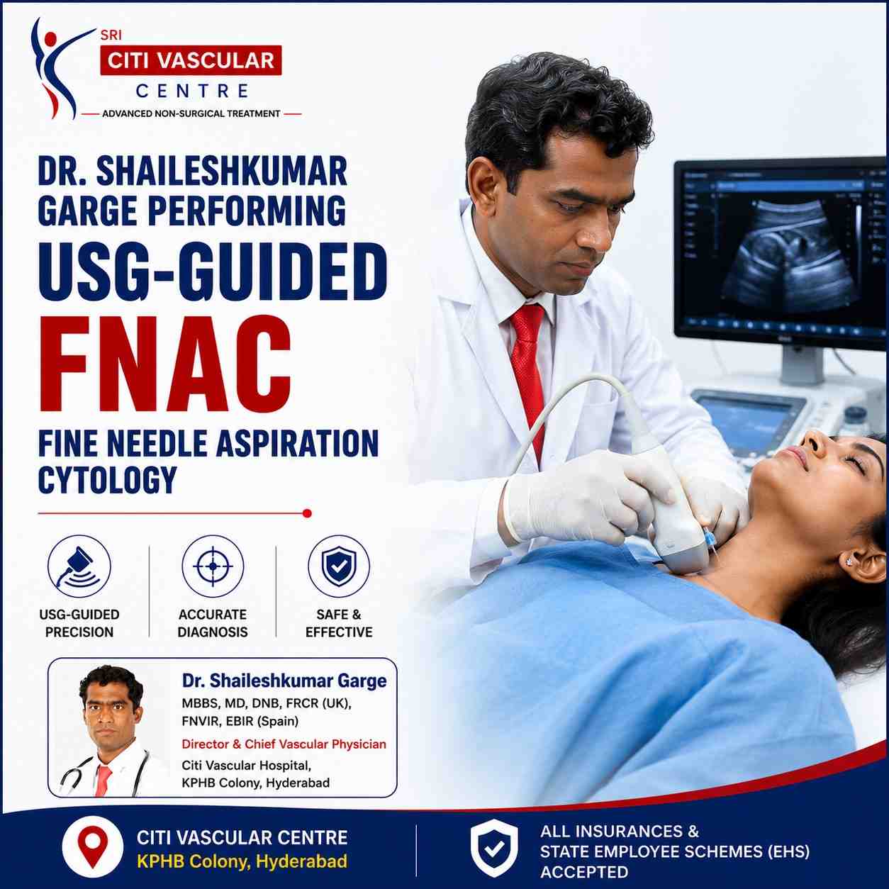
USG-Guided FNAC in Hyderabad — What You Need to Know
Fine needle aspiration under real-time ultrasound | 10-20 minutes | No stitches, no surgery | Same-day discharge | Diagnoses thyroid, breast, liver, lymph node, kidney, lung lesions | Local anaesthesia optional | Dr. Garge FRCR (UK) | Citi Vascular Centre, KPHB, Hyderabad
If your doctor has recommended an FNAC test, you probably have a lump, swelling, or abnormal area that showed up on an ultrasound or scan, and the next step is to find out what it actually is. USG-guided FNAC — Fine Needle Aspiration Cytology performed under real-time ultrasound guidance — is how that answer is obtained safely, quickly, and without surgery. A very fine needle is guided precisely into the lesion using continuous ultrasound imaging, and a small sample of cells is withdrawn for microscopic examination by a pathologist.
The procedure takes roughly 10 to 20 minutes, requires no stitches and no hospital admission, and most patients drive themselves home the same day. The ultrasound guidance is what makes modern FNAC fundamentally different from older palpation-based techniques — it allows the doctor to see exactly where the needle is going in real time, which is critical for small, deep, or difficult-to-feel lesions where a blind approach would risk missing the target entirely.
At Citi Vascular Centre, KPHB Colony, Hyderabad, USG-guided FNAC is performed by Dr. Shaileshkumar Garge — FRCR (UK), FNVIR (CMC Vellore), EBIR (Spain) — using advanced real-time imaging. This guide covers everything you need to know: what FNAC is and how it works, which organs can be assessed, what to expect on the day, how accurate it is, what the results mean, and when a biopsy might be needed instead.
Book Your FNAC Consultation — Citi Vascular Centre, KPHB, Hyderabad
Dr. Shaileshkumar Garge FRCR (UK) | Real-Time USG Guidance | Same-Day Discharge | Thyroid, Breast, Liver, Lymph Node, Kidney & More
Call +91-73375 83901 | WhatsApp | citivascularcentre.com
|
Feature |
Detail |
|
Full Form |
Fine Needle Aspiration Cytology — USG-Guided (ultrasound-guided) |
|
What Is Collected |
Cells (cytology) — not a tissue core. A fine hollow needle aspirates cells from the lesion. |
|
Imaging Used |
Real-time duplex ultrasound — needle position continuously visible on screen throughout the procedure |
|
Needle Size |
Very fine (21-25 gauge) — significantly thinner than a blood test needle |
|
Anaesthesia |
Usually NOT required. Local anaesthetic cream or injection offered for selected sites or anxious patients. |
|
Procedure Duration |
10-20 minutes for the actual sampling. Total visit including consultation and observation: 1-2 hours. |
|
Hospital Stay |
Day-care — no hospital admission required. Most patients go home within 1-2 hours of arrival. |
|
Stitches Required? |
No. The needle puncture seals naturally. No dressing except a small adhesive plaster. |
|
Pain Level |
Minimal — similar to a routine blood test. Most patients find it significantly less uncomfortable than expected. |
|
Organs Assessed |
Thyroid | Breast | Lymph nodes | Liver | Kidney | Lung (peripheral) | Salivary glands | Soft tissue |
|
Results Timeline |
Typically 2-5 working days — depending on the pathology laboratory |
|
Main Advantage |
Accurate cellular diagnosis of suspicious lumps without surgery — in most cases avoids the need for an operation |
When a scan or examination finds a lump, cyst, or abnormal area in the body, the next clinical question is always: what is it made of? Is it a simple benign cyst? A harmless tumour? An infection? Or something that needs urgent treatment? The answer requires looking at the actual cells inside that lesion — and that is exactly what FNAC does.
Fine Needle Aspiration Cytology is the process of inserting a thin hollow needle into a lesion and drawing cells into a syringe through gentle suction — much like taking a very localised blood sample, but from a tissue mass rather than a vein. The cells are placed on a glass slide, stained, and examined under a microscope by a pathologist, who can determine what type of cells are present and whether they appear normal, benign, inflammatory, or cancerous.
The 'USG-guided' part refers to ultrasound guidance — meaning the entire procedure is performed with real-time ultrasound imaging running continuously. The doctor watches the needle on the screen as it advances toward the target, confirming it is entering the correct part of the lesion. This is the critical difference from palpation-guided FNAC (where the doctor simply feels the lump and inserts the needle based on touch) — and it dramatically improves accuracy for small, deep, or difficult-to-palpate lesions.
FNAC vs Biopsy — The Key Difference
FNAC and biopsy are related but distinct procedures. FNAC collects individual cells (cytology) using a fine needle — quick, minimally invasive, and requiring no stitches. A core needle biopsy collects a small cylindrical core of tissue (histology) using a larger cutting needle — provides more structural information about the tissue architecture. FNAC is typically the first-line test because of its speed and minimal invasiveness. Biopsy is recommended when more tissue detail is required for definitive diagnosis or when FNAC is inconclusive.
FNAC is used whenever a clinical examination or imaging study has found an abnormality that needs cellular characterisation before treatment can be planned. Its primary value is answering the clinical question: what type of tissue is this?
|
Clinical Question |
What FNAC Determines |
|
Is this thyroid nodule cancerous or benign? |
Bethesda category — guides surgery vs active surveillance |
|
Is this breast lump a fibroadenoma or cancer? |
Distinguishes benign lesions (no surgery needed) from malignancy (immediate referral) |
|
Why are these lymph nodes enlarged? |
Identifies reactive swelling vs TB vs lymphoma vs metastatic cancer — each needs very different treatment |
|
Is this liver mass benign or malignant? |
Avoids laparotomy in many patients — directs targeted treatment |
|
What is this soft tissue lump? |
Lipoma vs cyst vs sarcoma — FNAC differentiates before surgery is planned |
Your doctor or specialist will recommend USG-guided FNAC when imaging studies or clinical examination have found an abnormality that requires a cellular diagnosis to plan appropriate management. FNAC is not a screening test — it is a targeted diagnostic procedure ordered when a specific finding needs characterisation
|
Finding |
Why FNAC Is Recommended |
|
Thyroid nodule > 1cm on ultrasound |
Standard first step before any thyroid surgery — avoids operation in benign nodules |
|
Breast lump or suspicious area on mammogram |
Triple assessment standard (clinical + imaging + FNAC) before any breast surgery |
|
Persistent enlarged lymph nodes |
Distinguishes reactive swelling, TB, lymphoma, and metastatic cancer without excision biopsy |
|
Liver or kidney mass on CT/MRI |
Tissue diagnosis before ablation or surgery — avoids major operation in many patients |
|
Salivary gland swelling |
Separates benign tumours (pleomorphic adenoma, Warthin) from carcinoma before surgery |
|
Soft tissue or subcutaneous lump |
Identifies benign lipoma/cyst vs sarcoma before excision planning |
|
Recurrent cyst requiring cytological analysis |
Drainage and cytology performed simultaneously — one procedure |
One of the great strengths of USG-guided FNAC is its versatility — it can be safely applied to a wide range of organs and anatomical sites throughout the body, provided they are accessible to the ultrasound beam. Here is a comprehensive guide to each organ site and what FNAC of that site is used for.
|
Organ |
Common Indications |
Clinical Notes |
|
Thyroid |
Thyroid nodules, goitre, suspected cancer |
Most common FNAC site. Results reported on Bethesda System (I–VI). |
|
Breast |
Breast lump, cyst, suspicious lesion on mammography/USG |
Part of triple assessment. Distinguishes fibroadenoma from carcinoma. |
|
Lymph Nodes |
Cervical, axillary, inguinal, retroperitoneal lymphadenopathy |
Critical: TB vs lymphoma vs metastatic — treatment completely different for each. |
|
Liver |
Hepatic mass, suspected metastases, HCC workup |
USG guidance avoids laparotomy. Accurate cellular diagnosis for most hepatic lesions. |
|
Kidney |
Renal mass with indeterminate radiological features |
Selected cases where cytology changes treatment planning before surgery or ablation. |
|
Lung |
Peripheral lung nodule close to chest wall |
USG-guided feasible for peripheral lesions; CT-guided preferred for central lesions. |
|
Salivary Glands |
Parotid and submandibular swellings |
Avoids diagnostic surgery for benign lesions. Identifies carcinomas requiring surgery. |
|
Soft Tissue |
Subcutaneous lumps, intramuscular masses, fluid collections |
Differentiates benign lesions from soft tissue sarcomas requiring urgent referral. |
Before ultrasound guidance became standard, FNAC relied entirely on the doctor feeling the lump and estimating needle position. For large, easy-to-feel lumps that was often adequate. For anything small, deep, complex, or near important structures, it was not. Real-time ultrasound changed this — the needle is visible on screen at every millimetre of its journey, making precise placement possible regardless of lesion depth or complexity.
|
Parameter |
Without USG Guidance |
With Real-Time USG Guidance |
|
Needle position |
Estimated by feel |
Confirmed on screen throughout |
|
Small lesions (< 1cm) |
High miss rate |
Reliably targetable — 5mm nodules sampled accurately |
|
Deep lesions (> 3cm) |
Very difficult — frequent inadequate samples |
Routinely accessible — depth measured on screen |
|
Vessel avoidance |
Cannot be seen |
Doppler identifies vessels before needle path is chosen |
|
Diagnostic yield |
Variable |
Significantly higher — operator confident in placement |
|
Complication risk |
Higher |
Lower — continuous visualization allows immediate adjustment |
The value of FNAC is that it can identify a wide spectrum of conditions from a single procedure. The pathologist who examines the collected cells can distinguish between the following categories, each of which requires very different clinical management.
|
Category |
Examples |
Management Impact |
|
Benign Tumours |
Fibroadenoma | Pleomorphic adenoma | Lipoma | Colloid nodule |
Reassurance — surgery usually avoidable. Surveillance plan offered. |
|
Malignant Tumours |
Thyroid papillary carcinoma | Breast carcinoma | HCC | Lymphoma | Metastatic cancer |
Urgent oncology or surgical referral. Staging investigations initiated promptly. |
|
Infectious / Inflammatory |
TB lymphadenitis | Granulomatous disease | Abscess | Reactive hyperplasia |
Appropriate antibiotics or anti-TB treatment. Avoids unnecessary surgery. |
|
Cystic Lesions |
Thyroid colloid cyst | Branchial cyst | Benign breast cyst |
Drainage and cytology simultaneously. Benign result avoids surgery. |
|
Suspicious / Indeterminate |
Bethesda III–IV thyroid | Atypical breast lesion |
Core biopsy or surgical excision recommended for definitive diagnosis. |
Much less than most patients expect. The needle used (21–25 gauge) is thinner than a standard blood test needle. Most people feel a brief sting for 2–3 seconds during insertion, mild pressure during aspiration, and then it is over. For thyroid, breast, and lymph node FNAC, no anaesthetic is needed at all. For deeper sites like liver or kidney, a small amount of local anaesthetic is injected at the skin surface first. Most patients leave saying it was easier than a blood test.
One of the practical advantages of FNAC over surgical biopsy is that preparation is simple. For most superficial sites — thyroid, breast, lymph nodes, salivary glands, and superficial soft tissue — no fasting and no medication changes are required. For selected deep abdominal procedures (liver, kidney, deep retroperitoneal lymph nodes), some specific preparation may be needed. Dr. Garge's team will provide site-specific instructions when you book your appointment.
|
When |
Step |
Notes |
|
At Booking |
Gather all previous imaging reports |
Ultrasound, CT, MRI, mammogram — helps Dr. Garge plan the optimal approach |
|
1 Week Before |
Inform team about blood-thinning medications |
Aspirin, clopidogrel, warfarin, apixaban, rivaroxaban — pause decision made per site |
|
Before Procedure |
Declare bleeding disorders and pregnancy |
Platelet count may be checked for deep organ FNAC |
|
Day of Procedure |
Fasting — generally NOT required |
Exception: deep abdominal FNAC (liver/kidney) may need 4–6 hour fast |
|
Day of Procedure |
Wear comfortable, accessible clothing |
Loose neckline for thyroid | front-open top for breast | separates for abdomen |
|
Transport |
Most patients can drive themselves home |
No sedation for standard FNAC — confirm if sedation planned for deep organ cases |
USG-guided FNAC is highly reliable when performed by an experienced operator using real-time imaging and interpreted by a qualified cytopathologist. Accuracy depends on six key factors:
|
Factor |
How It Is Optimised at Citi Vascular Centre, KPHB |
|
Ultrasound guidance quality |
High-resolution USG operated by Dr. Garge personally throughout — not delegated to a technician |
|
Operator experience |
12+ years dedicated interventional radiology | 15,000+ image-guided procedures |
|
Lesion characteristics |
Solid, viable component targeted — cystic or necrotic centre avoided to maximise cell yield |
|
Number of passes |
2–3 passes from different angles ensures adequate sampling from different lesion zones |
|
Sample adequacy check |
Visual assessment of slides before patient leaves — additional pass performed same session if needed |
|
Pathologist expertise |
Coordinated with experienced cytopathology partners for reliable, timely reporting |
Important Limitation: FNAC provides cellular (cytological) information. Some diagnoses requiring tissue architecture — follicular thyroid neoplasm, lymphoma sub-typing, liver fibrosis staging — may need a core needle biopsy instead. Dr. Garge will advise if this applies to your lesion.
Results are typically ready in 2–5 working days. Dr. Garge reviews every result with you at a follow-up appointment, correlating the cytology with your imaging and clinical findings. Results fall into five categories:
|
Result Category |
What It Means |
Common Examples |
Next Steps |
|
Benign |
No evidence of cancer or significant pathology |
Thyroid colloid nodule | Breast fibroadenoma | Reactive lymph node | Lipoma |
Reassurance. Surveillance plan discussed. Surgery usually not needed. |
|
Malignant |
Cancerous cells identified |
Thyroid papillary carcinoma | Breast carcinoma | Metastatic cancer | Lymphoma |
Urgent referral to oncology/surgery. Staging investigations. Treatment planning begins promptly. |
|
Suspicious |
Abnormal cells present but insufficient for a definitive diagnosis |
Bethesda III-IV thyroid | Atypical breast lesion | Follicular neoplasm |
Further evaluation recommended — repeat FNAC, core biopsy, or surgical excision biopsy |
|
Inflammatory |
Infection or inflammation rather than neoplasm |
Tuberculosis lymphadenitis | Abscess | Reactive hyperplasia | Granulomatous disease |
Appropriate antimicrobial or anti-inflammatory treatment. TB: CBNAAT/culture + anti-TB therapy. |
|
Inadequate / Unsatisfactory |
Sample does not contain enough cells for diagnosis |
Most common in highly fibrous lesions, very small nodules, or predominantly cystic lesions |
Repeat FNAC or core needle biopsy recommended. Not a normal finding — Dr. Garge will discuss next steps. |
FNAC results should never be interpreted in isolation. A benign cytology result in a clinically suspicious lesion still requires follow-up and possible further investigation. All results at Citi Vascular Centre are reviewed alongside your imaging and clinical picture before any management decision is made.
Recovery is one of FNAC's strongest practical advantages. Because the procedure involves only a fine needle puncture with no incision and no anaesthetic, most patients resume normal activities within hours. Here is what to expect:
|
Timeframe |
What to Expect |
Activity Guidance |
|
Immediately after |
20–30 min observation. Small plaster applied. Vital signs checked. |
Walk normally. Most patients drive home without assistance. |
|
First few hours |
Mild tenderness or pressure at puncture site. Possible small bruise. |
Normal daily activities. Avoid heavy manual work at the site. |
|
Day 1–2 |
Any soreness resolves. Bruise fades. |
Fully normal activity. Shower and remove plaster after 24 hours. |
|
Day 2–5 |
Await pathology result. No ongoing procedure symptoms expected. |
Normal life. Attend follow-up when Dr. Garge contacts with results. |
Call Citi Vascular Centre (+91-73375 83901) if you notice: significant swelling worsening over hours | pain not relieved by paracetamol | redness or discharge at the site | fever > 38°C. These are uncommon but should be assessed promptly.
USG-guided FNAC is one of the safest diagnostic procedures in interventional radiology. The combination of a fine needle and real-time ultrasound guidance that allows continuous visualization throughout keeps complication rates very low. Understanding the possible side effects — common but minor, and rare but more significant — helps patients have realistic expectations.
|
Complication |
Frequency |
Severity |
Management |
|
Mild pain at puncture site |
Very common |
Minimal |
Paracetamol/ibuprofen. Resolves within 24–48 hours. |
|
Small bruise |
Common |
Cosmetic only |
Self-resolving in 5–10 days. |
|
Temporary swelling |
Occasional |
Minimal |
Self-limiting. Cold compress if desired. |
|
Vasovagal faintness |
Uncommon — anxious patients |
Brief — momentary |
Procedure paused. Patient lies flat. Resolves immediately. |
|
Infection at site |
Rare (< 0.5%) |
Moderate if untreated |
Prevented by sterile technique. Short antibiotic course if confirmed. |
|
Significant bleeding |
Very rare with USG guidance |
Moderate if occurs |
Doppler identifies vessels pre-procedure. Direct compression usually sufficient. |
|
Pneumothorax (lung FNAC only) |
1–5% of peripheral lung FNAC |
Potentially significant |
Monitored post-procedure. Small pneumothorax often resolves spontaneously. |
|
No surgical incision |
Fine needle puncture seals naturally. No wound, no stitches, no scar. |
|
No general anaesthesia |
Local anaesthetic used only selectively — no intubation, no recovery room. |
|
Quick — 10–20 minutes |
Same-day discharge. Return to most activities the same afternoon. |
|
Avoids unnecessary surgery |
Benign result means surgery is often safely deferred or avoided entirely. |
|
Versatile — multiple organs |
Thyroid, breast, lymph node, liver, kidney, salivary gland — all under one roof at Citi Vascular, KPHB. |
|
Rapid diagnosis |
Result in 2–5 days vs surgical biopsy pathway (weeks). Treatment planning begins faster. |
|
Repeatable if needed |
Unlike surgery, FNAC can be safely repeated if the initial sample is inadequate. |
Patients Who Benefit Most from USG-Guided FNAC
USG-guided FNAC is appropriate for most patients presenting with an abnormal lump, mass, or swelling requiring cellular diagnosis. It is particularly well-suited to patients who: need a rapid diagnosis before planned surgery, have multiple lesions requiring simultaneous assessment, are anxious about surgery and want to know first whether it is actually needed, or have medical comorbidities that make surgical biopsy under general anaesthesia higher-risk.
|
Suitable Patients |
Why FNAC Is the Right First Test |
|
Thyroid nodule found on ultrasound |
FNAC is the standard diagnostic test before any thyroid surgery is considered. Surgery is avoided in benign nodules. |
|
Breast lump requiring diagnosis |
Triple assessment standard. FNAC confirms benign or malignant quickly — guides management decision. |
|
Persistent enlarged lymph nodes |
FNAC avoids diagnostic surgery (excision biopsy) in most cases — rapid access to cytological diagnosis. |
|
Suspected TB lymphadenitis |
FNAC confirms TB cytologically — enables starting anti-TB treatment without surgical excision |
|
Liver lesion on CT/MRI |
Avoids open or laparoscopic liver biopsy in most cases — FNAC provides hepatic cytology under ultrasound. |
|
Soft tissue lump of uncertain nature |
Lipoma vs cyst vs sarcoma — FNAC provides rapid answer before surgery is planned |
When FNAC May Not Be the Right Choice
|
Situation |
Why an Alternative Approach May Be Better |
|
Severe uncontrolled bleeding disorder |
Even a fine needle carries bleeding risk in anticoagulated patients. Platelet transfusion or correction of coagulopathy first. |
|
Diagnosis requires tissue architecture (histology) |
Follicular thyroid neoplasm, lymphoma sub-typing, most liver fibrosis — a core needle biopsy providing tissue cylinder is needed |
|
Patient unable to cooperate or remain still |
Movement during needle insertion risks inadequate sample or inadvertent injury — alternative approach or sedation |
|
Highly vascular lesion on Doppler |
For lesions with abundant feeding vessels, core biopsy with larger bore but haemostatic sheath may be safer |
|
Feature |
USG-Guided FNAC |
Palpation-Guided FNAC |
|
Needle position |
Confirmed on screen in real time |
Estimated based on feel |
|
Small lesions (< 1cm) |
Routinely achievable |
High miss rate |
|
Deep lesions |
Reliably accessible |
Very difficult |
|
Vessel avoidance |
Doppler identifies vessels before insertion |
Cannot be reliably avoided |
|
Diagnostic yield |
Significantly higher |
Variable — inadequate samples more frequent |
|
Best for |
All lesions — preferred technique in 2026 |
Only large, easily palpable, superficial lumps |
|
Differentiator |
Specific Evidence |
What This Means for Your Diagnosis |
|
Triple International Credentials |
FRCR (Royal College of Radiologists, UK) + FNVIR (CMC Vellore) + EBIR (European Board of IR, Spain) |
Internationally certified interventional radiologist — FNAC performed to international standards |
|
12+ Years Dedicated IR Experience |
Specialised exclusively in image-guided diagnostic and interventional procedures |
Not a general radiologist who 'also does' FNAC — image-guided procedures are his primary expertise |
|
Real-Time USG Guidance — Personal |
Dr. Garge performs the ultrasound guidance himself throughout every procedure — not delegated |
The same specialist who assesses your scan is the one who performs your FNAC — no handover |
|
15,000+ Minimally Invasive Procedures |
Total image-guided procedures at Citi Vascular Centre, KPHB |
High-volume operator — refined technique, consistent sample adequacy, low complication rates |
|
Full Findings Explanation |
Post-procedure consultation to explain what was sampled, what the pathology may show, and what happens next |
You leave understanding your procedure — not confused about what just happened and why |
|
One-Stop Imaging + FNAC |
Diagnostic ultrasound and FNAC performed in the same visit — no need for a separate imaging appointment first |
Single visit eliminates the typical two-appointment pathway (imaging then FNAC). |
|
Credential |
Detail |
|
Full Name |
Dr. Shaileshkumar Garge |
|
Qualifications |
MBBS | MD (Mumbai) | DNB (Delhi) | FRCR (UK) | FNVIR (CMC Vellore) | EBIR (Spain/Europe) | Fellowship (North Carolina, USA) |
|
Role |
Director and Chief Vascular Physician | Senior Consultant Vascular and Interventional Radiologist |
|
Centre |
Citi Vascular Centre, KPHB Colony, Road No. 1, Hyderabad, Telangana 500072 |
|
FNAC Sites |
Thyroid | Breast | Lymph Nodes | Liver | Kidney | Salivary Glands | Soft Tissue | Peripheral Lung | Fluid Collections |
|
Experience |
12+ years | 15,000+ image-guided diagnostic and interventional procedures |
|
What We Offer |
The Specifics |
Why It Matters |
|
Advanced Real-Time USG Equipment |
High-resolution ultrasound with colour Doppler — dedicated to diagnostic and interventional procedures |
Better image quality = better needle guidance = better sample adequacy |
|
One-Stop Diagnostic Visit |
Diagnostic USG assessment + FNAC in the same appointment — no second visit for the procedure |
Saves significant time and eliminates the anxiety of a second procedure appointment |
|
Day-Care Procedure — No Admission |
All FNAC procedures performed as outpatient day-care. No ward bed, no overnight stay. |
No disruption to work, family, or daily life |
|
Sterile Procedure Environment |
FNAC performed in a clean procedure room with full sterile precautions — skin prep, sterile draping, sterile needles |
Infection risk minimised by rigorous aseptic technique |
|
Coordinated Pathology Services |
Sample preparation and transport coordinated with experienced cytopathology laboratory |
Reliable sample handling and timely reporting |
|
Insurance Assistance |
Pre-authorisation support for medically indicated FNAC procedures where insurance coverage applies |
Reduces financial uncertainty |
|
Follow-Up Consultation |
Post-FNAC result review consultation with Dr. Garge to explain pathology and next steps |
No confusion about results — clear explanation of what was found and what it means for you |
|
1 |
Registration and review of previous imaging reports — ultrasound, CT, MRI, mammogram |
|
2 |
Consultation with Dr. Garge — explanation, consent, medication review, questions answered |
|
3 |
Pre-procedure ultrasound — fresh assessment of lesion, Doppler mapping of vessels, needle path planning |
|
4 |
USG-guided FNAC performed — 2–3 needle passes, watched on screen throughout. Most patients describe as equivalent to a blood test. |
|
5 |
Sample adequacy checked — if insufficient, additional pass performed in the same visit |
|
6 |
20–30 minute observation. Plaster applied. Vitals confirmed. |
|
7 |
Discharge with written instructions and contact number. Most patients drive home. |
|
8 |
Pathology result in 2–5 working days. Follow-up appointment with Dr. Garge to discuss findings and next steps. |
Q1: What is the purpose of a USG-guided FNAC test?
USG-guided FNAC collects cells from a lump or suspicious area using a fine needle guided by real-time ultrasound imaging. The cells are examined under a microscope to determine whether the lesion is benign, malignant, inflammatory, or infectious — providing a tissue diagnosis without surgery, and often avoiding an unnecessary operation in patients whose lesion turns out to be benign.
Q2: Is USG-guided FNAC better than a regular FNAC without ultrasound?
Yes — significantly better for most lesions. Real-time ultrasound guidance allows the needle position to be confirmed on screen throughout the procedure, which dramatically improves accuracy for small (< 1cm), deep, or complex lesions where palpation-guided FNAC would have a high miss rate and higher complication risk. At Citi Vascular Centre, KPHB, all suitable FNAC procedures use live ultrasound guidance.
Q3: Best FNAC centre near me in Hyderabad?
Citi Vascular Centre, KPHB Colony, Road No. 1, Kukatpally, Hyderabad — led by Dr. Shaileshkumar Garge FRCR (UK) — provides USG-guided FNAC for thyroid, breast, lymph nodes, liver, kidney, and soft tissue. Serving Kukatpally, Miyapur, Hitech City, Ameerpet, Secunderabad, and Telangana. Day-care procedure, same-day discharge. Mon–Sat 9AM–6PM. Call +91-73375 83901.
Q4: Which organs can undergo FNAC at Citi Vascular Centre, Hyderabad?
Dr. Garge performs USG-guided FNAC of the thyroid, breast, cervical and axillary lymph nodes, liver, kidney, salivary glands (parotid and submandibular), peripheral lung lesions, subcutaneous and soft tissue masses, and fluid collections at Citi Vascular Centre, KPHB. Virtually any lesion accessible to the ultrasound beam — including deep lesions not palpable on examination — can be sampled.
Q5: How long does FNAC take and when can I go home?
The needle sampling takes 10–20 minutes. Total clinic time — including consultation, pre-procedure ultrasound, the FNAC, and a 20–30 minute observation period — is typically 1.5–2 hours. No hospital admission is required. Most patients drive themselves home the same afternoon and resume normal daily activities the same day.
Q6: Can FNAC detect cancer?
Yes. FNAC can identify many cancers by examining the cellular characteristics of aspirated cells — thyroid papillary carcinoma, breast carcinoma, lymphoma, hepatocellular carcinoma, and metastatic disease among others. For some diagnoses requiring tissue architecture (follicular thyroid neoplasm, lymphoma sub-typing), a core needle biopsy providing a tissue cylinder may be needed in addition to or instead of FNAC.
Q7: When should I choose a core biopsy instead of FNAC?
FNAC is the preferred first-line test because it is quicker and less invasive. A core needle biopsy is recommended when FNAC is inconclusive, when the diagnosis requires tissue architecture rather than individual cells (follicular thyroid lesions, lymphoma sub-typing, liver fibrosis staging), or when Dr. Garge identifies a lesion where histological confirmation will directly change the management plan. Both options are available at Citi Vascular Centre, KPHB.
Q8: What happens if the FNAC report is inadequate?
An inadequate result means the sample did not contain enough diagnostic cells. Dr. Garge performs a visual adequacy check of the sample before you leave — if the initial passes are insufficient, an additional needle pass is made in the same visit. If the lesion is genuinely difficult to sample adequately (highly fibrous or predominantly cystic), a repeat FNAC or core needle biopsy will be recommended.
Q9: Do I need to fast before FNAC?
For most FNAC procedures — thyroid, breast, lymph nodes, salivary glands, and superficial soft tissue — no fasting is required. For deep abdominal FNAC (liver, kidney, retroperitoneal lymph nodes), fasting for 4–6 hours may be recommended for patient comfort and image quality. Dr. Garge's team confirms fasting requirements specific to your lesion site when you book your appointment.
Q10: How do I get my FNAC results?
Results are typically available in 2–5 working days. Dr. Garge contacts you and arranges a follow-up appointment to discuss the cytology report alongside your imaging and clinical findings. Results fall into five categories: Benign, Malignant, Suspicious (needs further testing), Inflammatory, or Inadequate. Results are never interpreted in isolation — clinical and imaging correlation is essential for every result category.
Q11: What are the risks of USG-guided FNAC?
USG-guided FNAC is one of the safest diagnostic procedures. Common minor side effects: mild tenderness and small bruise at the puncture site. Uncommon: momentary vasovagal faintness (managed immediately). Rare (< 0.5%): minor infection. Very rare: significant bleeding — Doppler identifies vessels before insertion. Pneumothorax is a specific risk with peripheral lung FNAC (1–5%) and is monitored post-procedure. Real-time ultrasound guidance minimises all risks.
Q12: Who is the best doctor for FNAC in Hyderabad?
Dr. Shaileshkumar Garge — FRCR (UK), FNVIR (CMC Vellore), EBIR (Spain) — Director and Chief Vascular Physician at Citi Vascular Centre, KPHB Colony, Hyderabad, is one of the city's most internationally credentialled interventional radiologists for image-guided FNAC. With 12+ years of dedicated interventional radiology and 15,000+ minimally invasive procedures, he performs USG-guided FNAC of all common sites. Call +91-73375 83901.
Citi Vascular Centre, KPHB Colony, Road No. 1, Hyderabad — USG-guided FNAC available for patients from:
Kukatpally and KPHB — 5 min
Miyapur and Bachupally — 10 min
Hitech City, Madhapur and Ameerpet — 20 min
Gachibowli and Kondapur — 25 min
Secunderabad and Begumpet — 25 min
Kompally, Medchal and Alwal — 20–25 min
Telangana and Andhra Pradesh — outstation welcome
|
Centre |
Contact |
Hours |
|
Citi Vascular Centre |
+91-73375 83901 |
KPHB Colony, Road No. 1, Hyderabad, Telangana 500072 | Mon–Sat 9AM–6PM |
|
Book Online |
citivascularcentre.com |
WhatsApp + online booking | Same-day FNAC available when scheduled | Insurance assisted |
|
USG-guided FNAC uses a very fine needle under real-time ultrasound to collect cells for microscopic diagnosis — no surgery, no stitches, no admission |
|
10–20 minutes procedure | Same-day discharge | Results in 2–5 days | Can be repeated if needed |
|
Accurate for thyroid, breast, lymph nodes, liver, kidney, salivary glands, and soft tissue masses |
|
Ultrasound guidance is essential for small (< 1cm), deep, or complex lesions — dramatically improves accuracy and safety |
|
Results: Benign | Malignant | Suspicious | Inflammatory | Inadequate — each with specific next steps discussed at follow-up |
|
A benign FNAC result avoids unnecessary surgery in many patients — one of the most significant clinical and economic benefits |
|
Dr. Shaileshkumar Garge FRCR (UK) | Citi Vascular Centre, KPHB | +91-73375 83901 | Mon–Sat 9AM–6PM |
When a scan finds something that needs explaining, USG-guided FNAC is how the answer is obtained quickly, safely, and without surgery. A very fine needle guided by real-time ultrasound aspirates cells from the lesion in 10 to 20 minutes. No stitches, no hospital bed, no general anaesthetic. The cytology result — ready in 2 to 5 working days — determines everything that follows: whether surgery is necessary, which type of cancer is present and needs urgent referral, or whether reassurance and surveillance are appropriate.
At Citi Vascular Centre, KPHB Colony, Hyderabad, Dr. Shaileshkumar Garge brings triple international credentials — FRCR (UK), FNVIR (CMC Vellore), EBIR (Spain) — and 15,000+ minimally invasive procedures to every FNAC consultation. The diagnostic ultrasound and FNAC are performed in the same visit, and every result is discussed personally with Dr. Garge at a follow-up appointment with imaging correlation.
Book Your USG-Guided FNAC
Thyroid | Breast | Lymph Node | Liver | Kidney | Salivary Gland | Soft Tissue
Dr. Shaileshkumar Garge | FRCR (UK) | FNVIR (CMC Vellore) | EBIR (Spain) | 12+ Years | 15,000+ Procedures
Call +91-73375 83901 | WhatsApp Now | citivascularcentre.com
Real-Time USG Guidance | Same-Day Discharge | No Stitches | Results in 2–5 Days | Insurance Assisted | Mon–Sat 9AM–6PM