LAST MEDICALLY REVIEWED:
July 2026 — Dr. Shaileshkumar Garge
Citi Vascular Hospital, KPHB Colony, Road No. 1, Hyderabad, Telangana 500072
QUICK ANSWER
How Is Prostate Artery Embolization Performed?
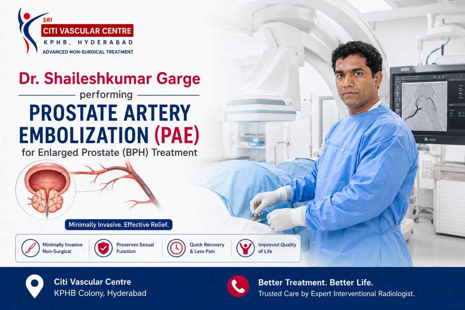
PAE is performed by an Interventional Radiologist through a 2mm puncture at the wrist or groin. A fine catheter is guided under real-time X-ray imaging (angiography) to the arteries supplying the prostate. Tiny microspheres are injected to reduce blood flow, causing the prostate to shrink gradually over 4–12 weeks. The procedure takes 60–90 minutes. Most patients go home the same day.
Prostate Artery Embolization (PAE) is a minimally invasive image-guided procedure used to treat benign prostatic hyperplasia (BPH) — the non-cancerous enlargement of the prostate that causes the urinary symptoms so many men live with for years before seeking help. Instead of removing enlarged prostate tissue through surgery — which is what TURP does — PAE works by interrupting the blood supply to the enlarged portions of the prostate. The prostate tissue that loses its blood supply gradually shrinks, the pressure on the urethra reduces, and urinary flow improves over the following weeks.
This page is the complete procedural guide to PAE — it explains every step of the preparation, the procedure itself, the access options, the imaging guidance used, what the recovery looks like day by day, the success rates in plain numbers, and what the risks and benefits are. If you are deciding whether PAE is right for you — or if you have already decided and want to know exactly what to expect — this is the page that answers all of it. For treatment overview and comparison, visit our main PAE page. For pricing and insurance, see our PAE Cost in Hyderabad page.
Book a PAE Consultation — Citi Vascular Centre, KPHB, Hyderabad
Call +91-73375 83901 | WhatsApp 73375 83901 | citivascularcentre.com | Mon–Sat 9AM–6PM
|
Feature |
Detail |
|
Procedure Type |
Minimally invasive catheter-based arterial embolization — no surgical incision |
|
Performed By |
Interventional Radiologist — specialised in image-guided catheter procedures |
|
Anaesthesia |
Local anaesthesia at wrist or groin + intravenous sedation. No general anaesthetic. |
|
Duration |
60–90 minutes for bilateral PAE. Complex anatomy may take 90–120 minutes. |
|
Access Point |
Radial artery (wrist) | Femoral artery (groin) |
|
Incision |
None — 2mm needle puncture only. No scalpel, no stitches required. |
|
Hospital Stay |
Day-care — most patients home same evening. Occasional overnight stay for selected cases. |
|
Catheter After Procedure |
Usually NOT required. Most patients void spontaneously post-PAE. |
|
Imaging Used |
Digital Subtraction Angiography (DSA) + Fluoroscopy + Cone-Beam CT (where required) |
|
Return to Desk Work |
7–10 days for most patients |
|
Return to Full Activity |
2–3 weeks |
|
Success Rate |
80–90% significant improvement in IPSS urinary symptom score at 1 year |
|
Prostate Volume Reduction |
20–40% reduction on MRI at 3–6 months |
|
First Symptom Improvement |
Most patients notice change at 4–6 weeks as prostate begins to shrink |
The term 'Prostate Artery Embolization' tells you exactly what the procedure does: it embolizes (blocks) the arteries (prostate arteries) of the prostate. The word embolization comes from the same root as embolus — a blocking agent — and in interventional radiology it refers to the deliberate, controlled introduction of material into an artery to reduce or stop blood flow to a specific tissue. In cancer treatment it is used to starve tumours. In fibroids it is used to shrink the uterine fibroid. In PAE, it is used to reduce blood flow to the enlarged, hyperplastic portions of the prostate.
What makes PAE technically distinct from other embolization procedures is the challenge of the target anatomy. The prostate artery is a small branch typically arising from the internal iliac artery — and it shares its blood supply neighbourhood with arteries to the bladder, rectum, and other pelvic structures that must absolutely NOT be embolized. The interventional radiologist must navigate a fine microcatheter precisely into the prostate artery — confirmed by angiographic imaging — before injecting the microspheres, ensuring they go where they are intended and not beyond. This level of selective targeting requires both advanced equipment and a high level of specialised experience.
PAE cannot be performed safely without real-time image guidance. The pelvic arterial anatomy is complex, varies between individuals, and the prostate arteries are often only 1–2mm in diameter — not visible to the naked eye and not accessible without navigating through a network of branching arteries from the groin or wrist. Here is what each imaging modality contributes to PAE safety and precision.
|
Imaging Modality |
What It Shows |
Why It Is Needed for PAE |
|
Digital Subtraction Angiography (DSA) |
Real-time X-ray images of arteries after contrast injection — background structures subtracted leaving only the vessels visible |
Shows prostate arterial anatomy in real time — confirms catheter position in the prostate artery before microspheres are injected |
|
Fluoroscopy |
Continuous live X-ray screening — shows catheter and wire position as they advance through the arterial system |
Allows real-time navigation from wrist or groin artery through to the prostate artery — the 'GPS' of catheter guidance |
|
Cone Beam CT (CBCT) |
3D rotational imaging of the prostate and its blood supply — provides CT-quality imaging in the angiography suite |
Confirms prostate artery selection, identifies non-target vessels, and verifies microsphere distribution before final embolization |
|
Road-Mapping |
DSA image overlaid on live fluoroscopy — creates a map of the arterial tree for catheter navigation |
Allows precise advancement of microcatheter through complex branching pelvic arteries without repeating contrast injections at every step |
PAE is performed exclusively by Interventional Radiologists — medical specialists who combine advanced training in diagnostic radiology with specific expertise in catheter-based minimally invasive procedures. Interventional Radiology is the specialty that performs image-guided procedures from within blood vessels, bile ducts, and other body channels — treating conditions that would otherwise require open surgery.
PAE is specifically performed by interventional radiologists because it requires: advanced catheter technique to navigate the pelvic arterial system safely, detailed knowledge of pelvic arterial anatomy and its variants, experience reading and interpreting angiographic imaging in real time, and the judgement to identify non-target vessels before injecting embolic material. At Citi Vascular Centre, KPHB, Hyderabad, PAE is performed by Dr. Shaileshkumar Garge — internationally trained Interventional Radiologist with FRCR (UK), EBIR (Europe), and FNVIR qualification in Hyderabad, with 12+ years of dedicated IR experience. For detailed specialist credentials, see our Best Doctor for PAE in Hyderabad page.
A well-prepared patient on the day of PAE leads to a smoother procedure, a more comfortable recovery, and better outcomes. Preparation begins at the consultation stage and involves specific clinical documents, tests, and lifestyle adjustments.
|
When |
Preparation Step |
Why |
|
At Consultation |
Bring prostate MRI (report + disc), PSA result, current medications list, previous prostate procedures history |
MRI maps prostate size and arterial supply — essential for PAE planning. PSA required to exclude cancer before BPH treatment. |
|
1–2 Weeks Before |
Prostate MRI (if not recently done) | Transrectal or transabdominal ultrasound (if available) |
MRI provides superior anatomical detail for PAE planning. Ultrasound confirms prostate size and post-void residual volume. |
|
1 Week Before |
Blood tests: full blood count, coagulation (PT/INR, APTT, platelets), renal function, PSA |
Coagulation screen essential before arterial access. Renal function confirms iodinated contrast can be safely administered. |
|
5–7 Days Before |
Blood thinners: aspirin usually continued. Warfarin and NOACs (apixaban, rivaroxaban) paused as per Doctors instruction |
Managing anticoagulation reduces bleeding risk at arterial puncture site without increasing thrombotic risk |
|
Night Before |
Fasting from midnight if sedation is planned — no food. Clear fluids until 4 hours before. |
Required for safe IV sedation administration |
|
Day Before |
Stop alpha-blockers (tamsulosin) the night before — as instructed |
Reduces post-procedure urinary side effects in some patients — specific to Dr. Garge's protocol |
|
Day of Procedure |
Comfortable loose clothing | Arrange a driver home | Bring all imaging discs and reports |
IV sedation means driving is not permitted on the day. Imaging needed on the day for reference. |
|
Before Start |
Written informed consent | IPSS questionnaire completed | IV cannula placed | Pre-procedure antibiotics given |
Antibiotics prevent post-PAE prostate infection (post-embolization syndrome). Consent confirms full understanding. |
The following is a complete step-by-step account of the PAE procedure as it is performed at Citi Vascular Centre, KPHB, Hyderabad. Understanding each step helps patients arrive prepared and comfortable — with no unexpected moments.
|
1 |
Arrival and Preparation You arrive at hospital and are taken to the preparation area. The team reviews your blood test results, confirms your allergy history, places an IV cannula in the back of your hand, and administers pre-procedure intravenous antibiotics. Your wrist or groin is shaved and cleaned with antiseptic. |
|
2 |
IV Sedation Administered Intravenous midazolam and fentanyl (or equivalent sedation agents) are given through the IV cannula. You remain conscious and can communicate throughout — but are deeply relaxed and comfortable. Most patients later describe the procedure as something they 'drifted through' rather than felt acutely. |
|
3 |
Local Anaesthetic and Arterial Access A small amount of local anaesthetic is injected at the wrist (radial artery) or groin (femoral artery) to numb the skin and deeper tissue. Once the area is fully numb, a very fine needle punctures the artery — this takes a fraction of a second. A tiny sheath (introducer) approximately 2mm in diameter is placed into the artery. You will feel pressure but no sharp pain. |
|
4 |
Catheter Guided to Pelvic Arteries A guidewire followed by a catheter is advanced through the sheath and navigated under continuous fluoroscopic X-ray guidance through the arterial system — from the wrist through the brachial and subclavian arteries into the aorta and down to the pelvic (internal iliac) arteries. The entire journey is visible on the screen throughout. |
|
5 |
Angiography — Mapping the Prostate Arteries A small amount of iodinated contrast dye is injected through the catheter while X-ray imaging runs continuously. This creates a real-time map (angiogram) of the arteries in the pelvis — allowing to identify the specific prostate arteries and their relationship to adjacent vessels supplying the bladder, rectum, and other pelvic structures. |
|
6 |
Microcatheter Positioned in Prostate Artery A hair-thin microcatheter is advanced through the larger catheter and positioned selectively inside the prostate artery — confirmed on angiography. Cone-beam CT may be used at this stage to confirm the microcatheter is positioned accurately within the prostate arterial territory and that no non-target vessels will receive microspheres. |
|
7 |
Microsphere Injection — First Side Calibrated microspheres (100–300 micron diameter) suspended in contrast solution are slowly injected through the microcatheter under continuous fluoroscopic monitoring. The flow of microspheres is watched on the screen — injection continues until the target level of flow reduction (stasis) is achieved in the prostate artery. This takes 5–15 minutes per side. |
|
8 |
Repeat on the Opposite Side (Bilateral PAE) The microcatheter is repositioned to the prostate artery on the opposite side — the procedure is repeated identically for the contralateral prostate artery. Bilateral (both sides) PAE is the standard approach, producing better and more durable outcomes than unilateral treatment in most patients. |
|
9 |
Final Angiography and Catheter Removal A final angiogram confirms satisfactory embolization of both prostate arteries with preserved flow to the adjacent bladder and rectal arteries. The microcatheter, catheter, and arterial sheath are removed. At the wrist, a compression band is applied for 2–3 hours. At the groin, firm pressure is held for 10–15 minutes. |
|
10 |
Transfer to Recovery — Observation You are transferred to the recovery area where nurses monitor your vital signs, pain level, and urinary function. Oral fluids and a light snack are offered when comfortable. Most patients can walk to the toilet within 1–2 hours. The observation period is typically 4–6 hours before discharge. |
The PAE procedure can be performed via two different arterial access points — the radial artery at the wrist or the femoral artery at the groin.
|
Feature |
Radial (Wrist) Access |
Femoral (Groin) Access |
|
Entry Point |
Radial artery — inner wrist |
Femoral artery — top of groin / thigh |
|
Compression After Procedure |
Small wrist compression band — 2–3 hours. Can sit and walk immediately. |
10–15 minutes manual pressure then bed rest for 2–4 hours to prevent bleeding at larger artery site |
|
Mobility Post-Procedure |
Can walk immediately after procedure. No restriction on sitting or standing. |
Bed rest for 2–4 hours at groin access site — minimises risk of haematoma at the puncture |
|
Bleeding Risk |
Lower — radial artery is smaller and more easily compressed |
Slightly higher — femoral artery is larger; haematoma at groin puncture site more common |
|
Patient Comfort |
Higher — patients can sit in a recliner chair during the observation period |
Lower — patients must lie flat for several hours post-procedure |
|
When Preferred |
When femoral artery is too small | When femoral artery spasm occurs | When radial arterial anatomy is more easily accessed from below |
First choice whenever anatomy and vessel size permit | Most PAE procedures at Citi Vascular Centre use this approach | |
The evidence base for PAE has grown substantially since its introduction — from early feasibility studies to large randomised controlled trials comparing it directly with TURP. Here is what the published data consistently shows.
|
Outcome |
Published Evidence |
|
Overall symptom improvement |
80–90% of patients achieve clinically significant improvement in IPSS score at 1 year. This is the headline result across multiple independent trials. |
|
Mean IPSS improvement |
8–15 point reduction in IPSS score — moving most patients from the severe category (score 20–35) into the moderate or mild range |
|
Prostate volume reduction |
20–40% reduction in prostate gland volume on MRI at 3–6 months |
|
Peak urinary flow rate |
Significant improvement in Qmax (maximum urinary flow rate) at 1 year — confirmed by UK-ROPE randomised controlled trial |
|
Nocturia improvement |
Most patients reduce nocturnal voids from 3–5 per night to 0–2 at 3 months — one of the most consistently reported early improvements |
|
5-year durability |
70–80% of patients maintain significant improvement at 5 years. Symptom recurrence is manageable with repeat PAE in most cases. |
For a complete discussion of PAE vs TURP outcomes, success rates at 2 and 5 years, and patient satisfaction scores — see our dedicated PAE vs TURP comparison page at citivascularcentre.com.
Clinical studies have shown that PAE provides significant improvement in urinary symptoms for many appropriately selected patients. Published studies have also demonstrated improvements in IPSS score, quality of life, urinary flow rate, and prostate volume while preserving sexual function in most men.
The first week after PAE is important for allowing the embolization to progress undisturbed and for avoiding complications at the arterial access site. Dr. Garge provides written discharge instructions with every PAE — but here is what every patient should expect to do and avoid in the days following their procedure.
|
Aftercare Area |
What to Do |
What to Avoid |
|
Wrist or Groin Site |
Keep dry and clean for 24 hours. Remove compression band after the time specifies. Small plaster until wound seals (1–2 days). |
Do not scratch, rub, or soak the puncture site. No baths for 24 hours — shower is fine. |
|
Medications |
Take prescribed antibiotics for full 7–10 days. Take anti-inflammatory medication (NSAID) as prescribed for 5–7 days — reduces post-PAE syndrome severity. |
Do not stop antibiotics early. Do not take other anti-inflammatory drugs without checking with Doctor. |
|
Hydration |
Drink 2–3 litres of water or clear fluids in the 24 hours after PAE — helps flush contrast dye through the kidneys. |
Avoid alcohol for 48 hours — increases dehydration and can interact with sedation residual effects. |
|
Physical Activity |
Walk normally from Day 1. Light activities from Day 3. Office work from Day 7–10. |
No heavy lifting (> 5kg) for 2 weeks. No strenuous exercise for 2 weeks. No swimming for 1 week. |
|
Driving |
Do not drive on the day of the procedure. Resume from Day 2 when sedation has fully cleared. |
Not on the day of PAE — sedation impairs reaction time. |
|
Urinary Symptoms |
Some worsening of urinary frequency and urgency in the first 1–4 weeks is expected and normal — this is the post-embolization inflammatory response. |
Do not stop BPH medications without instruction. Symptom worsening in the first weeks does not mean the PAE has not worked. |
|
Follow-Up |
Attend 6-week clinical follow-up. Complete new IPSS questionnaire at follow-up. |
Do not miss follow-up appointments — outcome assessment is essential for monitoring progress. |
Post-PAE Syndrome: Most PAE patients experience a flu-like period in the 24–72 hours after the procedure — low-grade fever, mild pelvic discomfort, fatigue, and sometimes nausea. This is a normal expected response to embolization of the prostate tissue and settles with the anti-inflammatory medication that is prescribed at discharge. It is not a sign that something has gone wrong. Call Us (+91-73375 83901) if your temperature exceeds 38.5°C, pain is not controlled by prescribed medications, or you cannot pass urine at all.
This is the question patients ask most often — and the answer is consistently better than most people expect. PAE is performed under local anaesthesia at the access site and intravenous sedation throughout, which means patients are relaxed, comfortable, and do not feel the catheter advancing through the arteries. The local anaesthetic at the wrist or groin numbs the puncture site completely before the needle is inserted.
During the procedure itself, patients typically feel nothing significant. Some patients notice mild warmth in the pelvic region when contrast dye is injected — a transient, expected sensation. A few patients experience brief pelvic cramping when microspheres are injected into the prostate arteries, but this settles immediately once injection pauses and is managed with the sedation already in place.
After the procedure, the most common discomfort is mild pelvic aching and the flu-like post-PAE syndrome described above — manageable with paracetamol and ibuprofen. Most patients rate day-of and day-after discomfort at 2–4 out of 10 — similar to a moderate period cramp — resolving within 48–72 hours. This is substantially less discomfort than recovering from TURP surgery, where catheter discomfort, surgical wound pain, and bladder spasms are common for the first week.
The PAE procedure at Citi Vascular Hospital, KPHB, typically takes 60 to 90 minutes from catheter insertion to catheter removal for bilateral PAE in patients with standard pelvic arterial anatomy. Complex cases — where the prostate arteries arise from unusual positions, where additional cone-beam CT is required to confirm targeting, or where the anatomy requires extra time for safe navigation — may take 90 to 120 minutes.
|
Time Component |
Duration |
|
Arrival, preparation and IV cannula |
30–45 minutes |
|
Sedation administration and preparation |
10–15 minutes |
|
Catheter insertion to prostate artery identification |
15–30 minutes |
|
Microsphere injection — first side |
10–20 minutes |
|
Repositioning and microsphere injection — second side |
10–20 minutes |
|
Final angiography and catheter removal |
10–15 minutes |
|
Recovery observation |
4–6 hours |
|
Total clinic visit time |
Approximately 6–8 hours from arrival to discharge for most patients |
Recovery from PAE is divided into two phases — the immediate post-procedure phase (hours to days) where the main focus is on the access site and post-embolization syndrome, and the gradual improvement phase (weeks to months) where the prostate progressively shrinks and urinary symptoms improve.
|
Timeframe |
Phase |
What to Expect |
|
Day 0 (Procedure Day) |
Observation and Discharge |
4–6 hours recovery. Vital signs monitored. Walk to toilet. Oral fluids and snack. Mild pelvic discomfort begins. Discharged home with antibiotics, anti-inflammatory, and written instructions. Wrist or groin compression band in place. |
|
Day 1–2 |
Post-PAE Syndrome |
Flu-like feeling — mild fever, pelvic aching, fatigue, occasional nausea. This is expected and normal. Manage with prescribed NSAID and paracetamol. Rest at home. Walk normally. Drink plenty of water. Do not drive. |
|
Day 3–5 |
Syndrome Resolving |
Fever and discomfort reduce significantly. Most patients feel markedly better by Day 3–4. Urinary frequency and urgency may still be slightly worse than before PAE — this is temporary inflammation, not failure. Light activities resumable. |
|
Day 7–10 |
Return to Light Work |
Most patients return to desk/office work. Driving resumable from Day 2 (once sedation fully cleared). Continue prescribed medications. Wrist site fully healed. Avoid heavy lifting. |
|
Week 2–3 |
Increasing Activity |
Urinary symptoms begin to stabilise and in many patients show first signs of improvement. Return to all normal activities except heavy gym work. Swimming permitted from Day 10–14. |
|
Week 4–6 |
First Symptom Improvement |
Most patients notice meaningful improvement in urinary flow, reduction in nocturia, and improved bladder emptying by 4–6 weeks. The prostate is shrinking. Complete IPSS questionnaire for comparison with baseline. |
|
Month 3 |
Follow-Up Assessment |
6-week (or 3-month) review with Dr. IPSS reassessed. Prostate MRI scheduled to confirm volume reduction. Most patients experiencing significant improvement. Some BPH medications may be reduced. |
|
Month 6 |
Maximum Benefit |
Prostate volume reduction at maximum level (20–40%). Most patients at their best urinary function post-PAE. IPSS at lowest level. Many patients stop BPH medications entirely with Dr’s guidance. 3–6 month outcome MRI reviewed. |
Important: Some patients experience paradoxical worsening of urinary symptoms in the first 2–4 weeks after PAE. This is due to temporary post-embolization inflammation and swelling of the prostate tissue — it resolves as the inflammation subsides and the prostate begins to shrink. Do not interpret early symptom worsening as PAE failure. Maximum benefit is typically seen at 3–6 months. Call the team on +91-73375 83901 if you are concerned.
|
No surgical incision |
2mm needle puncture only — no scalpel, no stitches, no scar at the procedure site |
|
No general anaesthesia |
Local anaesthesia and IV sedation only — safe for patients with cardiac, respiratory, or renal comorbidities where GA carries higher risk |
|
No catheter in most cases |
Most patients void spontaneously after PAE — the distressing post-surgical catheter experience is avoided |
|
Sexual function preserved |
Retrograde ejaculation — which affects 65–90% of TURP patients — occurs in fewer than 5% of PAE patients |
|
Same-day discharge for most patients |
No hospital ward admission in the standard protocol — home the same evening |
|
Minimal blood loss |
Catheter-based procedure — blood loss negligible compared to any surgical approach |
|
Quick return to normal activities |
Desk work from Day 7–10. Full activity at 2–3 weeks. Compare with TURP: 4–6 weeks |
|
Effective for very large prostates |
PAE works particularly well for very large prostates (> 80–100g) where surgical risk is higher and TURP is technically challenging |
|
Can be repeated |
Unlike surgery, PAE can be safely repeated if symptoms recur years later — in most cases without additional surgical risk |
• Men above 50 years
• Large prostate
• High surgical risk
• Want to preserve ejaculation
• Failed medicines
PAE is one of the safest BPH treatment procedures available — but like all medical interventions, it carries a defined risk profile. Understanding the possible complications helps patients make an informed decision and know what to watch for after the procedure.
|
Complication |
Frequency |
Severity |
Management |
|
Post-PAE Syndrome (flu-like) |
Very common — most patients |
Mild |
Expected and normal. Prescribed NSAID and paracetamol. Resolves within 48–72 hours. |
|
Bruising at wrist or groin |
Common |
Minimal — cosmetic |
Self-resolving within 7–10 days. Cold compress if tender. |
|
Temporary urinary worsening |
Common — first 2–4 weeks |
Temporary |
Expected inflammatory response. Settles as prostate shrinks. Contact team if acute retention. |
|
Arterial access site haematoma |
Uncommon |
Usually minor |
Compression and observation. Rarely requires intervention. |
|
Contrast dye reaction |
Rare (< 1%) |
Variable |
Managed in procedure suite with antihistamine, steroids. Severe allergy: 0.01% — pre-medication if prior history. |
|
Non-target embolization (bladder/rectum) |
Rare with experienced operator |
Can be significant |
Prevented by careful angiographic targeting and cone-beam CT confirmation. Managed if occurs. |
|
Prostate infection |
Rare (< 2%) |
Moderate if untreated |
Prevented by pre- and post-procedure antibiotics. Treated with antibiotics if confirmed. |
|
PAE failure (inadequate improvement) |
10–20% do not achieve adequate improvement |
No acute risk |
Repeat PAE or escalation to TURP — discussed at follow-up if improvement insufficient. |
|
Suitable for PAE |
Not Suitable for PAE — Consider Alternatives |
|
Moderate-severe BPH symptoms (IPSS > 8) inadequately controlled by medication |
Prostate cancer not yet excluded — PSA and urological review required first |
|
Sexually active men wanting to preserve ejaculatory function |
Active urinary tract infection — treat and resolve before any BPH intervention |
|
Very large prostate (> 80g) where TURP is technically challenging |
Bladder that has lost contractility — PAE relieves obstruction but cannot restore intrinsic bladder muscle |
|
High surgical risk — cardiac, respiratory, or renal disease precluding GA |
Severe renal impairment limiting safe contrast dye use |
|
Failed medical therapy (alpha-blockers, 5-ARIs) — seeking definitive treatment |
Neurogenic bladder from neurological disease — different management pathway required |
|
Men wanting to avoid hospital admission and return quickly to normal life |
Unsuitable pelvic arterial anatomy — rare; assessed on pre-procedure imaging review |
Suitability is determined at your PAE consultation after reviewing your prostate MRI, PSA, IPSS score, uroflowmetry results, and medical history. If PAE is not the right option for your specific anatomy or clinical picture, Dr. Garge will tell you clearly and coordinate an appropriate referral. Book your PAE consultation: +91-73375 83901 or WhatsApp 73375 83901.
Q1: How long does the PAE procedure take?
The PAE procedure itself takes 60–90 minutes for bilateral embolization in patients with standard pelvic arterial anatomy. Complex anatomy with unusual arterial variants may extend the procedure to 90–120 minutes. Total clinic time on the day — including arrival, preparation, IV sedation, the procedure, recovery observation, and discharge — is approximately 6–8 hours.
Q2: Is PAE painful?
PAE is far less painful than most patients expect. Local anaesthetic numbs the wrist or groin puncture site completely. Intravenous sedation keeps you deeply relaxed throughout. Most patients feel pressure but no sharp pain during the procedure. After the procedure, mild pelvic aching and flu-like symptoms lasting 24–72 hours are expected and managed with prescribed paracetamol and anti-inflammatory medication. Most patients rate discomfort at 2–4 out of 10.
Q3: Will I be awake during the PAE procedure?
Yes — PAE is performed under local anaesthesia and intravenous sedation, not general anaesthesia. You remain conscious and can communicate throughout, but the sedation makes you deeply relaxed and comfortable so that most patients drift through the procedure with minimal awareness. This is a significant advantage over TURP surgery — no intubation, no recovery from general anaesthesia, and no associated anaesthetic risks.
Q4: Will I need a catheter after PAE?
No — not in most cases. This is one of PAE's key advantages over TURP. Most patients void spontaneously after the procedure and do not require a catheter at any stage. A small minority — patients with pre-existing complete urinary retention or very large prostates — may need a temporary catheter for 24–48 hours post-procedure. Dr. Garge discusses the likelihood for your specific situation at your PAE consultation.
Q5: Can I eat and drink after the PAE procedure?
Yes — once you are comfortable in the recovery area, oral fluids are offered and light snacks are encouraged. Good hydration in the 24 hours after PAE is important to help flush contrast dye through the kidneys. There are no dietary restrictions after PAE. Avoid alcohol for 48 hours as it can interact with sedation residual effects and increase dehydration.
Q6: Will I need to stay in hospital after PAE?
Most PAE patients go home the same evening — day-care procedure. No ward admission is required in the standard protocol. A small number of patients — those with significant comorbidities, those who experience more than expected post-procedure discomfort, or those who do not void spontaneously before discharge — may stay overnight for observation. The Citi Vascular Centre team assesses every patient individually before discharge.
Q7: When can I drive after PAE?
Do not drive on the day of PAE — intravenous sedation impairs reaction time and judgement even after you feel fully alert. Most patients can drive from Day 2 onwards once the sedation has fully cleared. If you had a groin (femoral) access rather than wrist access, wait until the groin site is comfortable for the pedal pressure of driving — typically Day 3–5.
Q8: How soon will I notice improvement in urinary symptoms after PAE?
Improvement after PAE is gradual, not immediate — the prostate shrinks over weeks as the embolised tissue is reabsorbed. Most patients notice the first meaningful improvement at 4–6 weeks. Maximum benefit is typically reached at 3–6 months when prostate volume reduction is at its greatest. Some patients notice earlier improvement in nocturia (night-time waking) within 2–3 weeks of the procedure.
Q9: Is PAE done under X-ray? What imaging is used?
Yes — PAE is performed under continuous real-time X-ray imaging called fluoroscopy, combined with Digital Subtraction Angiography (DSA) which maps the prostate arteries by injecting contrast dye. Cone-Beam CT (a 3D imaging tool built into the angiography suite) may also be used to confirm accurate microsphere placement. The total X-ray exposure for PAE is comparable to a CT scan of the abdomen.
Q10: How long does it take for the prostate to shrink after PAE?
The prostate begins shrinking from the day of PAE as embolised tissue starts to be reabsorbed. The process is gradual — prostate volume reduces progressively over 3–6 months. Most patients experience 20–40% reduction in prostate volume at the 3–6 month MRI follow-up. Symptom improvement follows the same curve — earliest at 4–6 weeks, best results at 3–6 months.
Q11: Are there stitches after PAE?
No — PAE requires no stitches. The only entry into the body is a 2mm needle puncture at the wrist or groin. This seals naturally within 24 hours and requires nothing more than a small adhesive plaster. There is no wound to close, no dressing changes needed, and no stitch removal appointment. This is one of the practical advantages of PAE over any form of prostate surgery.
Q12: What is the success rate of PAE?
Published clinical trial data shows 80–90% of PAE patients achieve significant improvement in the International Prostate Symptom Score (IPSS) at 1 year. Mean IPSS score reduction is 8–15 points. Prostate volume reduces by 20–40% on MRI. Approximately 10–20% of patients do not achieve adequate improvement and may require repeat PAE or TURP. Dr. Garge discusses realistic expectations openly at your consultation. Call +91-73375 83901.
Citi Vascular Centre, KPHB Colony, Road No. 1, Hyderabad — PAE procedure for enlarged prostate available for patients from:
Kukatpally and KPHB — 5 min
Miyapur and Bachupally — 10 min
Hitech City, Madhapur and Ameerpet — 20 min
Gachibowli and Banjara Hills — 25 min
Secunderabad and Begumpet — 25 min
Kompally, Medchal and Alwal — 20–25 min
Telangana and Andhra Pradesh — outstation patients welcome
|
Centre |
Contact |
Hours |
|
Citi Vascular Centre |
+91-73375 83901 |
KPHB Colony, Road No. 1, Hyderabad, Telangana 500072 | Mon–Sat 9AM–6PM |
|
|
73375 83901 |
Share prostate MRI for initial PAE suitability assessment before booking |
PAE is performed by an Interventional Radiologist through a 2mm wrist or groin puncture — no surgery, no stitches, no general anaesthetic
The procedure takes 60–90 minutes. Total clinic time is 6–8 hours. Most patients home same evening.
10 clearly defined steps: arrival → sedation → access → catheter navigation → angiography → microcatheter placement → microspheres → bilateral repeat → final check → recovery
Wrist (radial) access is preferred — patients can walk immediately, more comfortable recovery, lower bleeding risk at access site
Post-PAE syndrome (flu-like, 24–72 hours) is expected and normal. Symptom improvement begins at 4–6 weeks. Maximum benefit at 3–6 months.
80–90% of patients achieve significant IPSS improvement at 1 year. 20–40% prostate volume reduction on MRI.
Citi Vascular Centre, KPHB | Dr. Garge FRCR (UK) | +91-73375 83901 | WhatsApp 73375 83901 | Mon–Sat 9AM–6PM
Prostate Artery Embolization is a 10-step catheter-based procedure performed under local anaesthesia and intravenous sedation — no surgery, no incision, no general anaesthetic, and no catheter for most patients. A 2mm puncture at the wrist gives Dr. Garge access to the arterial system. Under real-time fluoroscopic X-ray and angiographic guidance, a microcatheter is positioned precisely in the prostate arteries, and calibrated microspheres are injected to reduce blood flow. The prostate shrinks gradually over 4–12 weeks. 80–90% of patients achieve significant urinary symptom improvement at 1 year.
Recovery is straightforward — post-PAE syndrome for 24–72 hours, light activity from Day 3, office work from Day 7–10, and maximum benefit at 3–6 months. The procedure is performed at Citi Vascular Centre, KPHB Colony, Hyderabad, by Dr. Shaileshkumar Garge — FRCR (UK), FNVIR (CMC Vellore), EBIR (Spain) — one of Hyderabad's most internationally credentialled interventional radiologists. If you are considering PAE or want to know if you are a suitable candidate, WhatsApp your prostate MRI to 73375 83901 for an initial assessment before booking.
Book Your PAE Procedure Consultation — Citi Vascular Centre, KPHB, Hyderabad
PAE — No Surgery | No Catheter | No Sexual Side Effects | 60–90 Minutes | Same-Day Discharge
Dr. Shaileshkumar Garge | FRCR (UK) | FNVIR (CMC Vellore) | EBIR (Spain) | 12+ Years | 15,000+ Procedures
Call +91-73375 83901 | WhatsApp 73375 83901 | citivascularcentre.com
Mon–Sat 9AM–6PM | Insurance Assisted | 0% EMI Available | Transparent Pricing