LAST MEDICALLY REVIEWED:
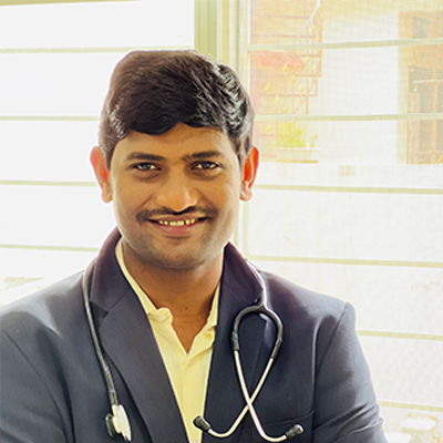
July 2026 — Dr. Shaileshkumar Garge
Citi Vascular Hospital, KPHB Colony, Road No. 1, Hyderabad, Telangana 500072
|
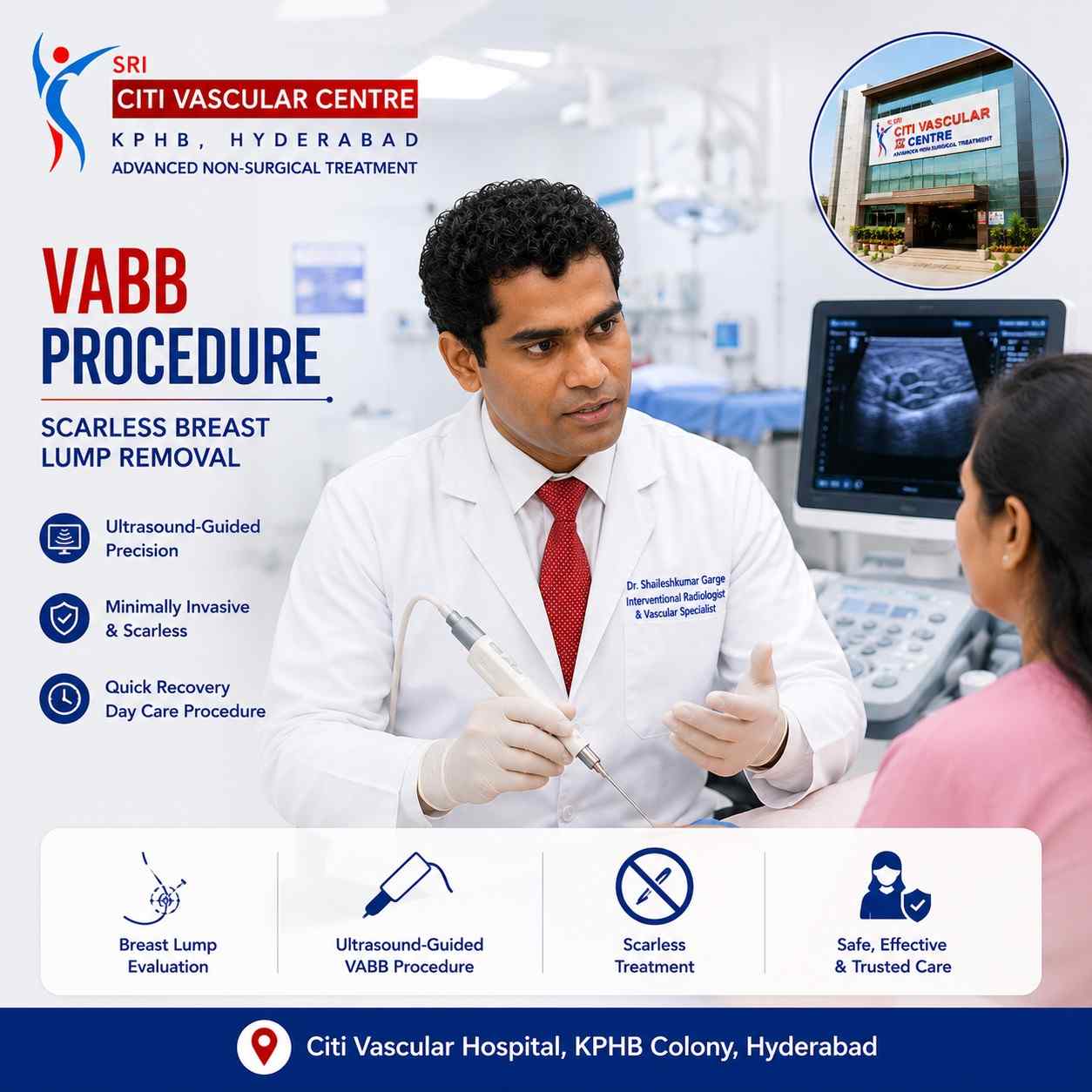
QUICK ANSWER What Is VABB and How Is It Used to Remove Breast Lumps Without Surgery? VABB (Vacuum-Assisted Breast Biopsy) is a minimally invasive, ultrasound-guided procedure that removes benign breast fibroadenomas through a 3–5mm skin nick under local anaesthesia. No surgical incision. No stitches in most cases. Same-day discharge. Recovery 1–3 days. Breast contour preserved with minimal or no visible scar. Performed by Dr. Garge FRCR (UK) at Citi Vascular Hospital, KPHB, Hyderabad. Call +91-73375 83901. |
For many women diagnosed with a breast fibroadenoma, the traditional path has been surgery — an operation under general or local anaesthesia, a visible incision in the breast, stitches, a recovery period of weeks, and a scar that may remain visible for life. For fibroadenomas that are small enough and confirmed benign on biopsy, this is not the only path available. Vacuum-Assisted Breast Biopsy — VABB — changes the equation entirely.
VABB was developed initially as a diagnostic technique to obtain larger tissue samples from breast lesions than standard needle biopsy could provide. Over time, as the technology evolved and operator experience grew, it became clear that VABB could do something even more useful for the right patient: completely remove a small benign fibroadenoma through a skin opening small enough to close without stitches in most cases — leaving essentially no surgical scar. The breast contour is preserved. The procedure takes 30 to 45 minutes under local anaesthesia. Most patients go home the same day and return to normal activities within a few days.
This page is the complete procedure guide for VABB at Citi Vascular Hospital, KPHB — covering the mechanism of action, the 10-step procedure, suitability, preparation, recovery day by day, risks, success rates, and cosmetic outcomes. For information about cost and insurance, see our VABB Cost in Hyderabad page. For a comparison of VABB versus conventional surgical excision, see our dedicated comparison page.
Book a VABB Consultation — Citi Vascular Hospital, KPHB, Hyderabad
Call +91-73375 83901 | WhatsApp 73375 83901 | citivascularcentre.com | Mon–Sat 9AM–6PM
|
Feature |
Detail |
|
Procedure |
Vacuum-Assisted Breast Biopsy (VABB) — minimally invasive image-guided tissue removal |
|
What It Treats |
Selected benign breast fibroadenomas and other benign breast lumps up to approximately 3–4cm |
|
Guidance Used |
Continuous real-time high-resolution ultrasound throughout the procedure |
|
Anaesthesia |
Local anaesthesia — skin and breast tissue numbed. No general anaesthetic required. |
|
Skin Opening |
3–5mm nick only — no surgical incision, no scalpel |
|
Stitches |
Not required in most cases — the small opening closes naturally within 24–48 hours |
|
Hospital Stay |
Day-care — home same day for most patients |
|
Procedure Duration |
30–45 minutes for most fibroadenomas. Multiple lesions may extend to 60 minutes. |
|
Recovery |
Mild soreness for 1–3 days. Desk work: Day 1–3. Full activity: Day 7–10. |
|
Scar |
Minimal or none — the tiny entry point fades to near-invisible in most patients |
|
Tissue Sent for Pathology? |
Yes — all removed tissue is sent for full histopathological examination as standard |
Vacuum-Assisted Breast Biopsy is an image-guided, minimally invasive technique that uses a specialised vacuum-assisted probe to remove breast tissue through a tiny skin opening under real-time ultrasound guidance. The procedure was originally developed as an advanced breast biopsy technique — designed to obtain larger, better-quality tissue samples than standard fine-needle or core needle biopsy could provide, without the need for open surgery. Over the past decade, the technique has evolved into a therapeutic tool: for selected patients with small confirmed-benign breast fibroadenomas, VABB can now completely excise the lump — not just sample it.
What makes VABB technically different from all other breast biopsy methods is the vacuum component. A standard core needle biopsy removes tissue passively — the needle cuts a small cylinder of tissue and withdraws it. VABB uses active negative pressure (suction) to draw breast tissue into the cutting aperture of the probe, then removes it in a controlled directional pass. By rotating the probe aperture systematically and repeating this process multiple times, the operator can progressively remove the entire volume of a small fibroadenoma in multiple passes — all through the same small skin entry point, under continuous ultrasound guidance that confirms complete removal in real time.
The critical distinction for patients is this: VABB is not the same as conventional breast surgery. There is no scalpel, no wound requiring closure, no drain, and no general anaesthetic. The procedure leaves a skin mark that is typically 3–5mm in diameter — smaller than many skin tags — which heals to near-invisibility over weeks to months. This is why VABB is classified as a scar-minimising, breast-preserving alternative to open excision for appropriately selected benign breast lesions.
The vacuum-assisted breast biopsy probe consists of a hollow outer needle with a side-cutting aperture and an inner rotating cutting mechanism connected to a vacuum pump. When the probe is positioned adjacent to the breast lump under ultrasound guidance, the vacuum draws the adjacent tissue into the cutting aperture — and the rotating cutting mechanism excises that tissue, which is then transported through the probe by suction to a collection chamber outside the breast. This single sequence — suction → tissue entry into aperture → cutting → removal → collection — takes a fraction of a second and is repeated systematically.
|
▼ |
Ultrasound localises the fibroadenoma precisely — size, depth, position relative to skin, distance from nipple, and proximity to any critical structures are all confirmed before any needle enters the breast |
|
▼ |
Local anaesthetic is infiltrated from skin surface down to the breast tissue level adjacent to the fibroadenoma — including the pericapsular tissue — numbing the area completely |
|
▼ |
A 3–5mm skin nick is made at the planned entry point using a small scalpel blade — this is the only 'cut' in the entire procedure |
|
▼ |
The VABB probe is introduced through the nick and advanced under continuous ultrasound guidance to the deep margin of the fibroadenoma — position confirmed before activation |
|
▼ |
Vacuum is applied — breast tissue is drawn into the aperture. The rotating cutter excises the tissue core, which travels through the probe to the external collection chamber |
|
▼ |
The aperture is rotated to the next position — the process repeats. Multiple passes are made systematically around and through the fibroadenoma until complete removal is confirmed on ultrasound |
|
▼ |
The ultrasound image is reviewed in real time throughout — the progressively disappearing fibroadenoma is visible as the echo-dense nodule reduces and then disappears from the imaging plane |
|
▼ |
All removed tissue cores are collected in the external chamber and are sent to the histopathology laboratory as standard — providing a complete tissue specimen regardless of whether the lump was already confirmed benign |
|
▼ |
A clip (small metallic marker) may be placed at the biopsy site to mark the location for future imaging reference — particularly useful for follow-up mammography or ultrasound |
|
▼ |
Firm pressure is applied to the breast for several minutes to stop any bleeding, then a compression dressing is applied. No stitches in most.. Patient observed for 30–60 minutes then discharged |
Patient selection is the most important factor in VABB outcomes — both for completeness of removal and for minimising complications. VABB is an excellent technique for the right patient and the right lump. Suitability is assessed at consultation by Dr. Garge after reviewing your breast ultrasound, BI-RADS category, FNAC or core biopsy result, and the lump's position within the breast.
|
Suitable Profile |
Clinical Basis |
|
Confirmed benign fibroadenoma on FNAC or core biopsy (Bethesda II) |
Benign histological or cytological confirmation is mandatory before VABB is planned for therapeutic (removal) rather than purely diagnostic purposes |
|
BI-RADS 2 or 3 on breast ultrasound |
Ultrasound features consistent with benign lesion — no suspicious features (irregular margins, microcalcifications, marked hypoechogenicity) |
|
Fibroadenoma size typically ≤ 3–4cm |
Most VABB devices work most effectively for fibroadenomas up to 3–4cm in maximum diameter. Some operators remove larger lumps with staged procedures. |
|
Growing benign fibroadenoma on serial ultrasound |
A confirmed-benign lump that has grown > 20% in two dimensions on follow-up — treatment indicated, VABB preferred over surgery in suitable size range |
|
Symptomatic fibroadenoma — pain, discomfort, or pressure |
Fibroadenomas causing breast discomfort or affecting quality of life despite confirmed benign histology warrant treatment — VABB when size permits |
|
Cosmetic concern — visible deformity or patient preference for removal |
A valid clinical indication when the lump creates visible breast asymmetry or the patient, after thorough counselling, prefers removal over continued monitoring |
|
Multiple small benign fibroadenomas in selected patients |
Two or more small benign fibroadenomas can sometimes be removed in the same VABB session — individually assessed at consultation |
|
Not Suitable When |
Clinical Reason / Alternative |
|
Suspected or confirmed breast malignancy |
VABB is not appropriate for suspected cancer — surgical wide local excision or mastectomy provides the margins and histological specimen required for definitive cancer treatment |
|
BI-RADS 4B, 4C, or 5 on breast imaging |
Intermediate to high suspicion for malignancy requires surgical excision providing complete specimen for definitive pathological staging |
|
Giant fibroadenoma (> 5cm) |
Very large fibroadenomas are generally beyond the reliable size range for complete VABB removal. Surgical excision provides more reliable complete removal. |
|
Rapidly growing lump raising suspicion of phyllodes tumour |
Phyllodes tumour — a rare but potentially aggressive breast tumour — requires wide surgical excision with adequate margins. VABB does not provide adequate margins. |
|
Uncorrected coagulopathy or active anticoagulation |
Haematoma risk at the biopsy site is higher with significant bleeding tendency. Anticoagulation must be managed before VABB is planned. |
|
Active breast infection or mastitis |
VABB is deferred until the infection is fully resolved with antibiotic treatment — introducing the probe through infected tissue increases haematoma and abscess risk |
|
Anatomy making safe probe access impossible |
Retroareolar (directly behind nipple) or very superficial lumps may carry higher risk of cosmetic or structural damage — assessed case-by-case |
|
No surgical incision |
3–5mm skin nick is the entire entry — no scalpel wound, no wound closure required, no wound care appointments |
|
Minimal or no visible scar |
The tiny entry point heals within 24–48 hours and fades to near-invisible over weeks. Compare with a surgical scar of 3–6cm after open excision. |
|
Local anaesthesia only |
No general anaesthetic required — safe for patients where general anaesthesia carries elevated risk due to comorbidities |
|
Same-day discharge |
No hospital ward admission. Most patients home within 2–3 hours of the procedure. Compare with open excision: day-case or overnight stay. |
|
Rapid return to normal activities |
Desk work from Day 1–3. Full activity by Day 7–10. Compare with surgical excision: 5–10 days for desk work, 2–3 weeks for full activity. |
|
Breast shape preserved |
No tissue defect from surgical excision — breast contour maintained. Particularly relevant for cosmetically sensitive areas. |
|
Simultaneous diagnosis and treatment |
All removed tissue is sent to histopathology — VABB provides a complete tissue specimen while simultaneously removing the lump |
|
Real-time confirmation of complete removal |
Ultrasound confirmation of complete lump excision before the probe is removed — the operator can see the fibroadenoma disappearing in real time |
|
Multiple lumps treated in one session |
Two or more small fibroadenomas can sometimes be treated in a single VABB procedure — avoiding multiple separate surgical operations |
|
Can be repeated |
If a new fibroadenoma develops in the same or opposite breast, VABB can be safely repeated — unlike surgery, which creates cumulative scar tissue |
Preparation for VABB is simpler than for any surgical procedure — but a systematic pre-procedure assessment is essential to confirm the lump is suitable for VABB and to plan the approach safely for your individual anatomy.
|
When |
Preparation Step |
Why |
|
At Consultation |
Breast ultrasound reviewed — BI-RADS, lump size, position, depth | Benign FNAC or core biopsy result confirmed |
Suitability assessment: size, location, and histological confirmation are all required before VABB is planned |
|
Before Procedure |
Blood tests if indicated: FBC, coagulation (PT/INR, platelets) — particularly for patients on blood thinners or with bleeding history |
Confirms haematoma risk is acceptable before needle insertion into breast tissue |
|
1–2 Weeks Before |
Blood thinners: aspirin usually continued. Warfarin and NOACs (apixaban, rivaroxaban) paused as per Dr. Garge's specific instruction |
Reduces haematoma risk at the VABB site without creating thromboembolic risk |
|
Day Before |
No special fasting required — VABB is under local anaesthesia only. Light meal on the day of procedure is appropriate. |
No general anaesthetic — strict fasting not required. Light meals reduce any discomfort during the procedure. |
|
Day of Procedure |
Wear comfortable, loose clothing with easy access to the chest area | Avoid perfume and deodorant on the breast | Bring all imaging discs and reports |
Sterile skin preparation is more effective on clean skin. Imaging needed for pre-procedure reference. |
|
Before Start |
Written informed consent | Pre-procedure breast ultrasound | IV cannula placed (safety precaution) | Local anaesthetic test dose |
IV cannula for safety in case emergency medication is needed. Test dose confirms absence of local anaesthetic allergy. |
The following is a complete account of the VABB procedure as performed at Citi Vascular Hospital, KPHB, Hyderabad. Understanding each step removes uncertainty and allows patients to arrive for their procedure relaxed and fully informed. Most VABB procedures take about 30–45 minutes, though the duration depends on the size, number, and location of the breast lesions.
|
1 |
Patient Positioning and Preparation You lie comfortably on your back with the arm on the affected side raised above the head — this position stretches and flattens the breast tissue, making the fibroadenoma easier to access and the procedure more comfortable. The breast is cleaned with antiseptic solution and sterile drapes are applied around the area. |
|
2 |
Pre-Procedure Planning Ultrasound Before any needle is introduced, Dr. Garge performs a detailed high-resolution ultrasound of the breast — confirming the fibroadenoma's exact position, depth below the skin surface, size in three dimensions, distance from the nipple and skin, and relationship to any important structures. The probe entry route is planned at this stage to maximise safety and efficiency. |
|
3 |
Local Anaesthetic Infiltration A fine needle is used to infiltrate local anaesthetic from the skin surface down to the breast tissue around and behind the fibroadenoma. The anaesthetic numbs the skin entry point, the breast tissue track, and the tissue surrounding the lump. This typically takes 3–5 minutes to reach full effect. Most patients feel the initial skin prick and mild pressure — after which the area is completely numb. |
|
4 |
Tiny Skin Nick Once the local anaesthetic has taken full effect, a small scalpel blade makes a 3–5mm skin nick at the planned entry point. This is the only true incision in the procedure — and it is smaller than a standard ear piercing. This nick allows the VABB probe to pass into the breast tissue without resistance. |
|
5 |
VABB Probe Insertion Under Ultrasound The vacuum-assisted probe is introduced through the skin nick and advanced under continuous real-time ultrasound guidance to the deep (far) margin of the fibroadenoma. Position is confirmed on the ultrasound screen before any activation of the device — the probe tip is clearly visible as a bright echo within the breast tissue adjacent to the lump. |
|
6 |
Vacuum-Assisted Tissue Removal — First Pass The vacuum is activated: negative pressure draws adjacent fibroadenoma tissue into the side aperture of the probe. The rotating cutter fires, excising the tissue core, which travels through the probe by suction to the collection chamber. A single pass takes a fraction of a second. Dr. Garge watches the ultrasound throughout — the tissue being removed is visible in real time. |
|
7 |
Systematic Coverage — Multiple Passes The probe aperture is rotated to the next position (typically in 30-degree increments) and the vacuum-cut cycle repeats. This is done systematically around the entire circumference of the fibroadenoma — ensuring complete coverage of the lump volume. During this phase, the ultrasound shows the fibroadenoma progressively reducing as tissue is removed. |
|
8 |
Real-Time Ultrasound Confirmation of Complete Removal When all passes are complete and the fibroadenoma is no longer visible on ultrasound, a completion scan confirms that the lump has been entirely removed. If any residual fibroadenoma tissue is detected, additional targeted passes are performed. The operator can see the biopsy cavity — a small hypoechoic (dark) space where the lump once was. |
|
9 |
Clip Placement and Probe Removal A small metallic clip marker (approximately 2mm) may be placed at the biopsy site before the probe is removed — this marks the location for future mammography or ultrasound reference. The probe is then withdrawn through the same 3–5mm nick. Firm pressure is applied to the breast for 5–10 minutes to minimise any bleeding. |
|
10 |
Compression Dressing and Observation A firm compression dressing (not a surgical wound dressing — simply an adhesive pad with firm elastic support) is applied to the breast. You are moved to a comfortable chair in the observation area. Vital signs are checked. Most patients feel quite well at this stage. After 30–60 minutes of observation confirming no significant haematoma, you are discharged with written aftercare instructions and a follow-up appointment. |
VABB is performed under local anaesthesia — which means you remain fully conscious and alert throughout the entire procedure. There is no intubation, no general anaesthetic, no operating theatre, and no anaesthetist required. Local anaesthetic (typically lidocaine with adrenaline, which also reduces local bleeding) is injected into the skin and breast tissue before the procedure begins.
Most patients experience the initial injection of local anaesthetic as mild discomfort — comparable to a dental injection — followed by complete numbness of the breast in the treated area within 3–5 minutes. Once the area is numb, most patients feel pressure and movement during the procedure but no pain. The vacuum-cut cycle itself produces a clicking sound and a sensation of pulling — neither of which is painful once the local anaesthetic is fully active.
Intravenous sedation (midazolam or equivalent) can be added for patients who feel particularly anxious, at the treating clinician's discretion — this produces deep relaxation while maintaining consciousness. Most patients who have VABB without sedation describe the experience as very manageable — often less anxiety-provoking than they anticipated before the procedure.
Can I drive home after VABB? If the procedure was performed under local anaesthesia alone — yes, you can drive yourself home. If IV sedation was administered, you cannot drive on the day and must arrange a driver. Confirm this with the team at booking so you can plan appropriately. Call +91-73375 83901 or WhatsApp 73375 83901.
|
Timeframe |
Phase |
What to Expect |
|
Day 0 (Procedure Day) |
Discharge |
30–60 min observation. Light snack and drink. Compression dressing in place. Mild breast soreness beginning. Home same day. Avoid heavy lifting. Rest comfortably on back avoiding pressure on breasts. |
|
Day 1 |
Immediate Recovery |
Mild to moderate breast soreness — paracetamol adequate. Compression dressing maintained. You can shower (keeping dressing dry). Light daily activities normal. Most patients manage with paracetamol only. |
|
Days 2–3 |
Settling |
Soreness settling significantly. Some bruising around the biopsy site is common — appears purple/yellow over 5–7 days, then fades. Desk and office work resumable from Day 2–3 for most patients. |
|
Days 4–7 |
Normal Activities |
All normal daily activities resumable. Light exercise (walking) from Day 3–4. Avoid heavy gym work and contact sports for the first 2 weeks. Compression dressing can usually be replaced with a supportive sports bra. Can take shower. |
|
Week 2 |
Bruising Resolving |
Bruising substantially resolved. Small skin nick healing — looks like a minor skin mark at this stage. Histopathology result of removed tissue typically available by Day 5–10. Dr. Garge contacts you with the result. |
|
Weeks 3–4 |
Cosmetic Healing |
Skin nick fading — typically reduced to a pale mark of 3–5mm. Full activity including gym resumable. Post-procedure clinic review with Dr. Garge at 4–6 weeks to discuss histology result and follow-up schedule. |
|
Months 1–3 |
Scar Maturation |
Entry point continues fading — most patients describe the mark as imperceptible or indistinguishable from normal skin texture at 3 months. First follow-up ultrasound confirms biopsy cavity has resolved and no residual lump. |
|
Area |
What to Do |
What to Avoid |
|
Compression Dressing |
Keep in place and dry for 24 hours. Replace with a firm supportive sports bra from Day 2. |
Do not remove the compression dressing prematurely on Day 0. Do not soak in bath until Day 3–4. |
|
Skin Nick |
No dressing change needed in most cases — the small nick seals naturally. Shower from Day 1 (covering the area with a waterproof dressing if available). |
Do not rub or scratch the healing site. No creams or topical preparations on the nick for 48 hours. |
|
Pain Management |
Paracetamol 500–1000mg up to 4 times daily as needed. NSAID (ibuprofen) if prescribed. |
Do not take aspirin for pain (increases bruising). Do not exceed maximum paracetamol dose. |
|
Physical Activity |
Light walking from Day 1. Desk work from Day 2–3. Driving from Day 1 (if no sedation was given). |
No heavy lifting (> 5kg) for 2 weeks. No vigorous upper body exercise for 2 weeks. No swimming for 1 week. |
|
Bra Support |
Wear a firm, supportive sports bra (no underwire) continuously for the first 3–5 days — day and night. This reduces haematoma risk significantly. |
Avoid underwired bras for 1 week after the procedure. Avoid sports bras that compress uncomfortably. |
|
When to Call Urgently |
Call +91-73375 83901 immediately if: rapidly increasing breast swelling | breast becomes very hard and painful | temperature > 38°C | skin redness spreading from the site |
None of these are expected — these are uncommon but require prompt review if they occur |
VABB has a very favourable safety profile — particularly when performed by an experienced operator under continuous ultrasound guidance. The absence of a surgical incision, the avoidance of general anaesthesia, and the localised nature of the procedure all contribute to a complication rate that is substantially lower than open surgical excision. The following table presents the documented complications divided by frequency.
|
Complication |
Frequency |
Severity |
Management |
|
Bruising around biopsy site |
Very common |
Cosmetic only |
Self-resolving within 7–14 days. Cold compress in first 24 hours may reduce extent. No treatment required. |
|
Mild breast soreness |
Very common — Day 1–3 |
Mild |
Paracetamol and NSAID adequate. Firm bra support reduces discomfort significantly. |
|
Small haematoma (blood collection) |
Uncommon — 2–5% |
Minor in most cases |
Firm compression and observation. Most resolve spontaneously within 2–3 weeks. Very rarely requires aspiration. |
|
Vasovagal episode (brief faint) |
Uncommon — < 5% |
Transient |
Brief light-headedness related to local anaesthetic or anxiety. Patient positioned flat, recovers within minutes. Prevented by IV cannula access. |
|
Wound site infection |
Rare — < 1% |
Manageable |
Short antibiotic course if confirmed. Sterile technique prevents in most cases. Watch for increasing redness and discharge. |
|
Incomplete fibroadenoma removal |
5–15% for larger fibroadenomas |
Residual lump — not dangerous |
Detected by post-procedure and follow-up ultrasound. Managed with repeat VABB or observation depending on residual size. |
|
New fibroadenoma elsewhere in breast |
Occurs over time — not related to VABB technique |
Benign |
Detected on follow-up ultrasound. Same management pathway as the original lump — VABB if appropriate. |
|
Significant haematoma requiring drainage |
Very rare — < 0.5% |
More significant |
Aspiration or surgical drainage if the haematoma is expanding or not resolving. Very uncommon with appropriate post-procedure compression. |
Published evidence for VABB in the therapeutic removal of benign breast fibroadenomas is extensive — with large series from Korea, Italy, China, and European centres consistently reporting high technical success rates and excellent patient satisfaction with cosmetic outcomes.
|
Outcome |
Published Evidence and Clinical Expectation |
|
Complete fibroadenoma removal at single session |
Technical success (complete removal confirmed on post-procedure ultrasound) in 85–95% of appropriately selected patients with fibroadenomas ≤ 3cm. Higher residual rate for larger fibroadenomas. |
|
Symptom improvement |
Cosmetic concern, palpable lump, and breast discomfort all resolve with complete removal. Most patients notice immediate absence of the palpable lump after the procedure. |
|
Patient satisfaction with cosmetic outcome |
High — studies consistently report 85–95% patient satisfaction with the cosmetic result. The tiny skin nick is the main determinant. Most patients cannot identify the entry site at 3 months. |
|
Histopathological confirmation rate |
100% — all removed tissue is sent to histopathology as standard, providing definitive histological diagnosis of every lump removed by VABB |
|
Residual fibroadenoma requiring repeat procedure |
Approximately 5–15% of patients require a second VABB session for incomplete removal — most commonly with larger fibroadenomas (3–4cm). Most are managed with observation if residual tissue is very small. |
|
New fibroadenoma development at any site |
Fibroadenomas can develop de novo in breast tissue that was not part of the original lump — this is the natural history of fibroadenoma tendency, not a VABB failure. Managed on their own merits at follow-up. |
For a detailed comparison of VABB outcomes versus conventional surgical excision — including recovery time, scarring, recurrence rates, and patient-reported satisfaction scores — see our dedicated VABB vs Surgery comparison page at citivascularcentre.com.
|
Credential / Facility |
Relevance to VABB for Breast Fibroadenoma |
|
FRCR (UK) — Royal College of Radiologists |
Highest UK postgraduate radiology standard — covers expert breast imaging (BI-RADS) and interventional breast procedures including VABB |
|
FNVIR (CMC Vellore) |
India's most prestigious IR fellowship — direct training in image-guided breast procedures including USG-guided FNAC, core biopsy, and vacuum-assisted breast biopsy |
|
EBIR (Spain/Europe) |
European Board of Interventional Radiology — international certification encompassing European guideline-level breast intervention standards |
|
Personal ultrasound operation throughout VABB |
Dr. Garge operates the high-resolution ultrasound personally throughout every VABB procedure — probe positioning, tissue removal, and completion confirmation all by the same specialist |
|
Real-time ultrasound confirmation of complete removal |
The most important technical safeguard in VABB — confirms complete fibroadenoma removal before the probe is withdrawn. Reduces residual lump rate. |
|
Histopathology sent as standard for every VABB |
All tissue removed by VABB is sent for full histopathological examination — not assumed benign based on pre-procedure FNAC alone. Complete diagnostic + therapeutic in one procedure |
|
Written pre-procedure estimate |
Transparent pricing before any commitment. Insurance pre-authorisation assisted. 0% EMI for eligible patients. See VABB Cost page for full pricing detail. |
|
Credential |
Detail |
|
Name |
Dr. Shaileshkumar Garge | MBBS | MD (Mumbai) | DNB (Delhi) | FRCR (UK) | FNVIR (CMC Vellore) | EBIR (Spain) | Fellowship (North Carolina, USA) |
|
Role |
Director and Chief Vascular Physician | Senior Consultant Vascular and Interventional Radiologist |
|
Centre |
Citi Vascular Hospital, KPHB Colony, Road No. 1, Hyderabad, Telangana 500072 |
|
Experience |
12+ years dedicated interventional radiology | 15,000+ minimally invasive image-guided procedures |
The information on this page is based on current principles of minimally invasive breast interventions and image-guided breast procedures. Vacuum-Assisted Breast Biopsy (VABB) is an established technique used worldwide for the diagnosis and selected therapeutic removal of benign breast lesions. Treatment recommendations should always be individualized after clinical examination and breast imaging by a qualified specialist.
Key Clinical References
American College of Radiology (ACR) – ACR Appropriateness Criteria®: Palpable Breast Masses and Breast Imaging guidelines for the evaluation and management of breast lesions.
Society of Interventional Radiology (SIR) – Clinical practice guidance supporting image-guided minimally invasive procedures and best practices for patient safety and quality care.
European Society of Breast Imaging (EUSOBI) – Evidence-based recommendations on breast imaging, image-guided biopsy techniques, and multidisciplinary management of breast lesions.
National Institute for Health and Care Excellence (NICE), UK – Guidance on the diagnosis, assessment, referral, and management of breast conditions and suspected breast cancer where applicable.
Peer-reviewed Medical Literature – Recommendations are supported by published evidence in internationally recognized journals, including studies on ultrasound-guided Vacuum-Assisted Breast Biopsy, fibroadenoma management, cosmetic outcomes, complication rates, and patient satisfaction.
Q1: What is Vacuum-Assisted Breast Biopsy (VABB)?
VABB is a minimally invasive, ultrasound-guided procedure that uses a vacuum-assisted probe introduced through a 3–5mm skin nick under local anaesthesia to remove benign breast fibroadenomas without conventional surgery. The procedure combines real-time ultrasound guidance with negative pressure technology to systematically remove the lump in multiple passes through the same small entry point. No surgical incision. No stitches in most cases. Same-day discharge at Citi Vascular Hospital, KPHB.
Q2: Is VABB painful?
VABB is performed under local anaesthesia — the skin and surrounding breast tissue are numbed before the probe is introduced. Most patients experience the initial local anaesthetic injection as mild discomfort, after which the breast is completely numb. During the procedure, patients feel pressure and movement — but no pain. Post-procedure soreness for 1–3 days is common and managed comfortably with paracetamol. Most patients rate the overall experience at 2–3 out of 10.
Q3: Is VABB a surgery?
No — VABB is specifically classified as a minimally invasive procedure, not surgery. It requires no operating theatre, no surgical incision, no general anaesthesia, and no surgical team. The only skin opening is a 3–5mm nick that requires no stitches in most cases. Recovery is measured in days rather than weeks. VABB is performed in a dedicated procedure room, not an operating theatre, by an Interventional Radiologist rather than a surgeon.
Q4: Can VABB completely remove a fibroadenoma?
Yes — complete removal of the fibroadenoma is confirmed by real-time ultrasound at the end of every VABB procedure at Citi Vascular Hospital. Published data shows technical success (complete removal on post-procedure imaging) in 85–95% of appropriately selected patients with fibroadenomas ≤ 3cm. A small proportion — approximately 5–15%, more common with larger lumps — may have residual tissue detected on follow-up ultrasound, which is managed with repeat VABB or observation.
Q5: Will I have a scar after VABB?
Minimal or no visible scar. The only entry into the breast is through a 3–5mm skin nick — no surgical wound, no stitches required in most cases. This tiny entry point heals within 24–48 hours and fades progressively over weeks. Most patients cannot identify the entry site at 3 months. This is one of the most valued outcomes of VABB — particularly compared with the 3–6cm surgical scar that remains after conventional open excision.
Q6: Can I go home the same day as VABB?
Yes — VABB is a day-care procedure. After 30–60 minutes of observation confirming no significant haematoma, most patients are discharged home. No hospital ward admission. No IV drip, no drain, no urinary catheter. You will need a driver if IV sedation was given — but if VABB was performed under local anaesthesia only, you can drive yourself home. The team at Citi Vascular Hospital confirms discharge requirements at booking. Call +91-73375 83901.
Q7: When can I return to work after VABB?
Most patients return to desk or office work within 1–3 days of VABB. Light physical work from Day 3–5. Heavy manual work and vigorous gym exercise are avoided for 2 weeks to reduce haematoma risk. Swimming is deferred for 1 week until the skin entry point is fully sealed. Driving is possible from Day 1 if no sedation was given. The specific return-to-work timeline depends on your occupation — discuss with Dr. Garge at your consultation.
Q8: Is VABB safe? What are the risks?
VABB has a very favourable safety profile when performed by an experienced operator under ultrasound guidance. The most common side effects are bruising (very common, self-resolving within 1–2 weeks) and mild soreness for 1–3 days. Haematoma (blood collection at the biopsy site) occurs in approximately 2–5% of cases and usually resolves without intervention. Significant complications are uncommon. Serious complications such as infection or injury to adjacent structures are rare (< 1%).
Q9: Can a fibroadenoma come back after VABB?
The removed fibroadenoma does not grow back — tissue that is removed is gone. However, the breast tissue that produced the original fibroadenoma may produce new fibroadenomas elsewhere over time — this is the natural history of the individual's tendency to form fibroadenomas, not a VABB failure. Approximately 5–15% of patients develop residual or new fibroadenomas requiring attention within 12 months. Long-term ultrasound follow-up detects these early.
Q10: Will the removed tissue be sent for laboratory examination?
Yes — all tissue removed during VABB is sent to histopathology as standard at Citi Vascular Hospital, KPHB. This provides a definitive histological diagnosis of the lump — confirming benign fibroadenoma and excluding any unexpected finding such as a phyllodes tumour or atypia that may not have been apparent on pre-procedure FNAC. Results are typically available within 5–10 days. Dr. Garge contacts you with the result and reviews it at your follow-up appointment.
Q11: Who is the best doctor for VABB in Hyderabad?
Dr. Shaileshkumar Garge — FRCR (UK), FNVIR (CMC Vellore), EBIR (Spain) — Director and Chief Vascular Physician at Citi Vascular Hospital, KPHB Colony, is one of Hyderabad's most internationally credentialled interventional radiologists for breast fibroadenoma evaluation and VABB scarless removal. With 12+ years of dedicated IR experience and 15,000+ image-guided procedures, he performs VABB with real-time ultrasound guidance and histopathology as standard. Call +91-73375 83901.
Q12: Which hospital offers VABB for breast fibroadenoma in Hyderabad?
Citi Vascular Hospital, KPHB Colony, Road No. 1, Hyderabad, led by Dr. Shaileshkumar Garge — FRCR (UK), FNVIR (CMC Vellore), EBIR (Spain) — provides complete breast fibroadenoma evaluation and VABB scarless removal: high-resolution breast ultrasound, USG-guided FNAC, core biopsy, and VABB with same-day discharge, real-time completion confirmation, and standard histopathology. Insurance assisted. Transparent pricing. Call +91-73375 83901 or WhatsApp 73375 83901.
Citi Vascular Hospital, KPHB Colony, Hyderabad — VABB scarless breast lump removal for patients from:
Kukatpally and KPHB — 5 min
Miyapur and Bachupally — 10 min
Hitech City, Madhapur and Ameerpet — 20 min
Gachibowli, Kondapur and Banjara Hills — 25 min
Secunderabad and Begumpet — 25 min
Kompally, Medchal and Alwal — 20–25 min
Telangana and Andhra Pradesh — outstation patients welcome
|
Centre |
Contact |
Hours |
|
Citi Vascular Hospital |
+91-73375 83901 |
KPHB Colony, Road No. 1, Hyderabad, Telangana 500072 | Mon–Sat 9AM–6PM |
|
|
73375 83901 |
Send breast ultrasound and FNAC report for initial VABB suitability assessment | Same-week appointments | Insurance assisted |
|
VABB removes benign breast fibroadenomas through a 3–5mm skin nick under local anaesthesia and real-time ultrasound — no surgical incision, no stitches, no general anaesthetic |
|
10-step procedure: positioning → planning scan → local anaesthetic → skin nick → probe insertion → vacuum-cut (multiple passes) → ultrasound confirmation → clip placement → compression → observation + discharge |
|
Same-day discharge. Soreness 1–3 days. Desk work Day 1–3. Full activity Day 7–10. Entry point fades to near-invisible at 3 months. |
|
All removed tissue sent to histopathology as standard — complete diagnostic and therapeutic procedure in one visit |
|
Technical success (complete removal) in 85–95% of appropriately selected patients ≤ 3cm. 85–95% patient satisfaction with cosmetic outcome. |
|
Benign FNAC (Bethesda II) and BI-RADS 2–3 on ultrasound are mandatory prerequisites before VABB is scheduled for therapeutic removal |
|
Dr. Shaileshkumar Garge FRCR (UK) | Citi Vascular Hospital, KPHB | +91-73375 83901 | WhatsApp 73375 83901 | Mon–Sat 9AM–6PM |
Vacuum-Assisted Breast Biopsy is one of the most patient-friendly advances in breast care of the past decade. For women with small confirmed-benign breast fibroadenomas who want their lump removed — without a surgical scar, without general anaesthesia, and without weeks of recovery — VABB is the evidence-based minimally invasive alternative. A 3–5mm skin nick, 30 to 45 minutes under local anaesthesia, real-time ultrasound confirmation of complete removal, and full histopathology on every removed specimen. Most patients go home the same day and return to normal life within days.
The procedure is not appropriate for every breast lump — confirmed benign histology and appropriate size on ultrasound are prerequisites, and certain lump characteristics or patient factors make surgery the more appropriate recommendation. VABB in the right patient is an outstandingly effective, cosmetically excellent, and clinically safe procedure. For an assessment of whether VABB is right for your specific fibroadenoma — WhatsApp your breast ultrasound report and FNAC result to 73375 83901, or call +91-73375 83901 to book a consultation at Citi Vascular Hospital, KPHB Colony, Hyderabad.
VABB — Breast Lump Removal. No Surgery. No Scar. Same Day Home.
3–5mm Skin Nick | Local Anaesthesia | 30–45 Minutes | Same-Day Discharge | Breast Contour Preserved
Dr. Shaileshkumar Garge | FRCR (UK) | FNVIR (CMC Vellore) | EBIR (Spain) | 12+ Years | 15,000+ Procedures
Call +91-73375 83901 | WhatsApp 73375 83901 | citivascularcentre.com
Transparent Pricing | Insurance Assisted | 0% EMI | Citi Vascular Hospital, KPHB | Mon–Sat 9AM–6PM