LAST MEDICALLY REVIEWED:
August 2026 — Dr. Shaileshkumar Garge
Citi Vascular Hospital, KPHB Colony, Road No. 1, Hyderabad, Telangana 500072
QUICK ANSWER
Who Is the Best Doctor for Osteoid Osteoma Treatment in Hyderabad?
Osteoid osteoma treatment may involve CT-guided percutaneous ablation or surgical excision, depending on the lesion's location, imaging findings and individual clinical requirements. An Interventional Radiologist experienced in CT-guided bone lesion ablation can assess suitability for procedures such as radiofrequency ablation (RFA) and other appropriate techniques. Treatment selection should be based on clinical assessment and discussion of the available options. Call +91-73375 83901.
Osteoid osteoma is a benign bone lesion that commonly affects children, adolescents and young adults and may cause pain that worsens at night. Treatment options include CT-guided percutaneous ablation and surgical excision. The appropriate approach depends on the diagnosis, lesion location, accessibility, patient factors and the clinical judgement of the treating team.
This page is not a repetition of the clinical content on our Osteoid Osteoma Ablation Procedure, RFA, Cost, or Surgery vs Ablation pages. Those pages explain what the treatments involve. This page answers the different and equally important question: how do you evaluate whether a specific doctor and hospital are the right choice, and what should you look for — and ask — before committing to a treatment that in many cases involves general anaesthesia, significant cost, and a recovery period that affects school and sports for weeks?
Book an Osteoid Osteoma Consultation — Citi Vascular Centre, KPHB, Hyderabad
WhatsApp CT scan to 73375 83901 | Call +91-73375 83901 | Mon–Sat 9AM–6PM
Book your Appointment here: | Appoinment: |Call +91-7032508468| Whatsapp:+91-7337583901
|
Consideration |
What to Look For |
|
Primary Specialist |
Interventional Radiologist with specific CT-guided bone lesion ablation training — not a general radiologist performing occasional ablation |
|
Essential Qualifications |
Review the practitioner's relevant qualifications, recognised specialist training and experience in CT-guided bone lesion procedures. Verify qualifications and professional registration through the appropriate official sources. |
|
CT Interpretation |
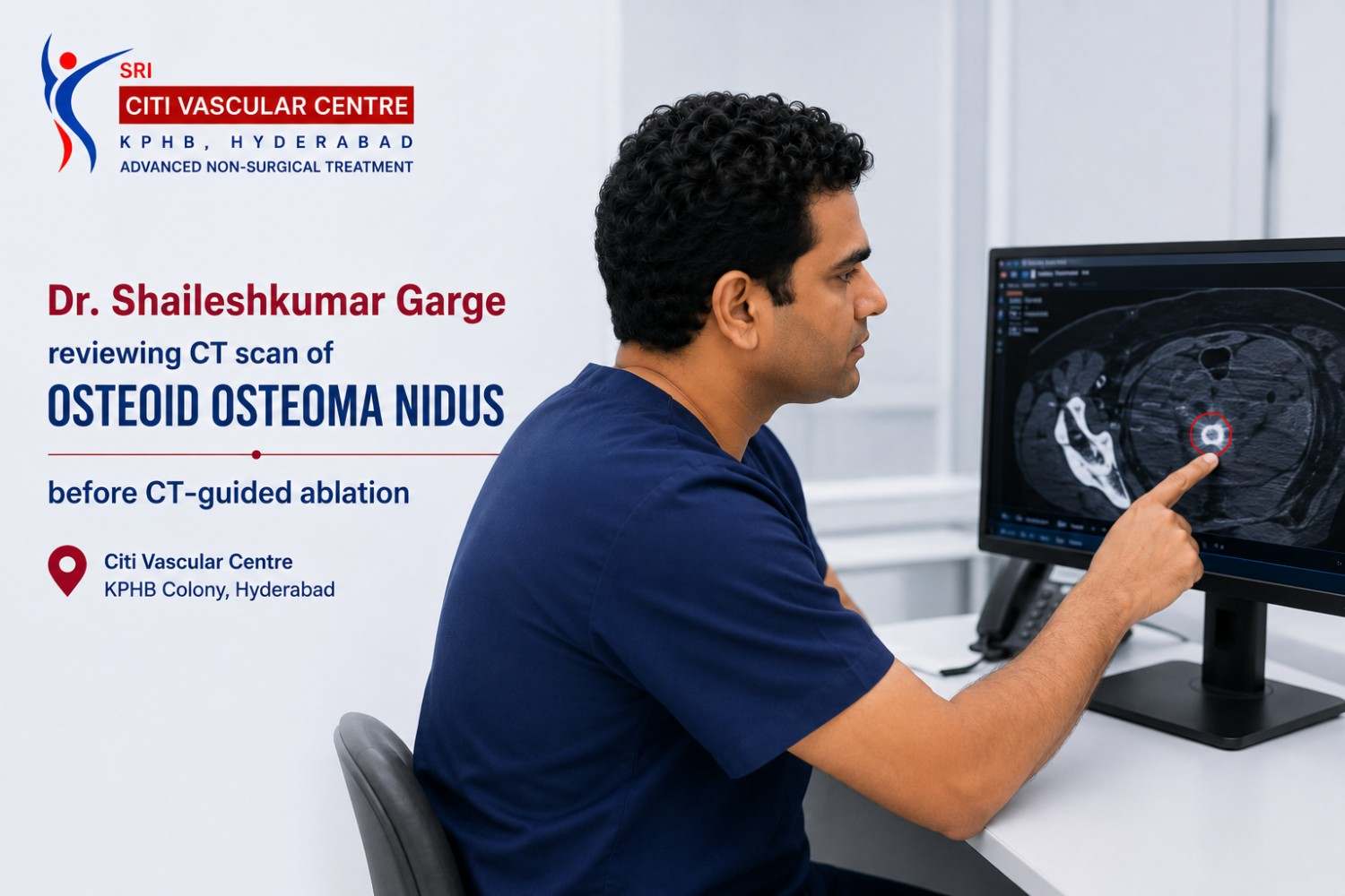
Must review the thin-slice CT personally — identifying the nidus, measuring depth and proximity to nerves, planning the access route. Not delegate to a technician. |
|
Ablation Technique Range |
Available ablation techniques include radiofrequency ablation, microwave ablation and cryoablation. The most appropriate technique depends on lesion location, proximity to adjacent structures, equipment availability and the treating team's assessment.. |
|
Diagnostic Rigour |
Should confirm CT diagnosis before planning ablation — not treat without clear nidus visualisation. Should advise biopsy when diagnosis is uncertain. |
|
Paediatric Experience |
OO most often affects children and teenagers — the specialist should be experienced with GA-based paediatric ablation, not exclusively adult patients. |
|
Transparent Communication |
Written cost estimates | honest discussion of repeat treatment likelihood | specific recovery guidance for your child's sport and school schedule |
CT-guided osteoid osteoma ablation requires accurate identification of the nidus, appropriate needle positioning and careful selection of the ablation technique. Planning should consider the lesion's size, depth, anatomical location and proximity to nerves, joints and surrounding structures.
Experience with CT-guided bone lesion procedures is relevant when evaluating a practitioner for osteoid osteoma treatment. Patients may ask about the practitioner's training, experience with similar lesion locations, approach to procedural safety and management of complications.
The consequence of the wrong choice of specialist is real: a missed nidus (because the CT was not carefully reviewed and the planned entry trajectory did not reach the target), an incomplete ablation (because the probe tip was not confirmed within the nidus before energy delivery), a nerve injury (because a heat-based ablation was used without hydrodissection adjacent to a named nerve), or an inappropriate recommendation for surgery when ablation was technically achievable. The right specialist makes the right assessment before any procedure begins.
Red flag: Before ablation is planned, the treating practitioner should review the relevant imaging, assess whether the nidus can be identified confidently and explain the proposed access route and potential risks. Additional investigation or an alternative treatment may be appropriate when the diagnosis or lesion location is uncertain.Call +91-73375 83901 if you have concerns about a prior assessment.
A patient or parent searching for osteoid osteoma treatment in Hyderabad will encounter several different specialist types. Understanding what each does — and what their specific limitations are — helps navigate the referral pathway effectively.
|
Specialist |
Role in OO Management |
Key Consideration |
|
Interventional Radiologist (IR) |
CT-guided percutaneous thermal ablation — RFA, microwave, cryoablation | CT nidus review | Treatment planning | Paediatric and adult ablation |
An Interventional Radiologist with appropriate experience may evaluate patients for CT-guided percutaneous ablation. Suitability depends on the imaging findings, lesion location and individual clinical requirements. |
|
Orthopaedic Surgeon (general) |
Surgical excision of OO — open or arthroscopic | Bone biopsy | Structural management |
Appropriate when: diagnosis is uncertain (histology needed) | percutaneous access impossible | patient prefers excision. Some orthopaedic surgeons have limited experience with ablation as an alternative — confirm whether they can refer for ablation if appropriate. |
|
Orthopaedic Oncologist |
Specialists in bone tumours — both benign and malignant. OO falls within their scope for diagnosis and surgical management. |
Particularly relevant when the diagnosis is not yet confirmed or when a malignant bone lesion needs to be excluded. May refer to IR for ablation if OO diagnosis is established. |
|
Paediatric Orthopaedic Surgeon |
Surgical OO management in children — limb-specific expertise for growth plate considerations in growing bone |
Relevant for very young children where growth plate proximity is a significant concern affecting treatment planning. Works with paediatric IR for ablation cases in children. |
|
Paediatric IR |
CT-guided ablation specifically in children — paediatric GA, dose-reduction CT protocols, child-appropriate probe sizing |
Relevant for very young children where paediatric-specific IR expertise is needed. Dr. Garge treats paediatric OO patients at Citi Vascular Centre with paediatric anaesthetic collaboration. |
For patients considering minimally invasive osteoid osteoma treatment in Hyderabad, consultation with an appropriately qualified practitioner experienced in CT-guided bone lesion ablation may help determine the available treatment options. Orthopaedic assessment may also be appropriate when the diagnosis is uncertain or surgical management is being considered.Dr. Garge at Citi Vascular Centre, KPHB, provides this assessment, performs all ablation modalities, and refers to orthopaedic surgery when that is the more appropriate recommendation for your specific case. Call +91-73375 83901.
|
1 |
Personal CT Nidus Review |
The specialist must personally review the thin-slice CT (ideally 1–2mm slice CT) and specifically identify the nidus — its exact location within the bone, its depth below the skin, its proximity to adjacent nerves or joint surfaces, and the planned needle access route. A specialist who plans treatment from a standard radiology report without reviewing the CT images themselves is working with incomplete information that significantly increases the risk of a misplaced probe tip and ablation failure. |
|
2 |
Full Ablation Technique Range |
Different ablation techniques may be suitable for different osteoid osteoma locations. Cryoablation or additional protective techniques may be considered in selected cases near nerves or joint structures. The treating team should explain why a particular technique is appropriate and discuss surgical alternatives when necessary. |
|
3 |
Diagnostic Honesty — Will Recommend Surgery When Appropriate |
Treatment recommendations should be based on the diagnosis, lesion characteristics and clinical suitability. When ablation is not appropriate, the practitioner should discuss further investigation, referral or alternative treatment options. |
|
4 |
Paediatric Competence |
Because osteoid osteoma most commonly affects children, teenagers, and young adults — the specialist must be experienced in managing paediatric patients: GA rather than sedation in young children, paediatric radiation dose reduction CT protocols, growth plate awareness in treatment planning, and post-procedure guidance for school, sports, and physical education that reflects the realities of a child's daily life rather than adult workplace guidelines. |
|
5 |
Transparent, Specific Communication |
Patients and caregivers should receive clear information about the proposed procedure, estimated costs, anaesthesia requirements, potential complications, follow-up arrangements and activity restrictions. Recovery recommendations should be individualised according to the lesion location, treatment performed and the patient's clinical progress. |
|
Q |
Ask the Specialist |
What a Good Answer Looks Like |
|
1 |
Have you personally reviewed my CT scan — and can you describe the nidus position to me? |
The practitioner should explain the nidus location, relevant imaging findings, proposed needle access route and proximity to important anatomical structures, including any uncertainties or procedural risks. |
|
2 |
Is the diagnosis confirmed — or do you need to do a biopsy first? |
An honest answer: if the CT features are classical (lucent nidus, surrounding sclerosis, clinical night pain), ablation without biopsy is appropriate. If CT features are atypical, the answer should be 'we need to confirm the diagnosis first.' |
|
3 |
What ablation technique are you planning — RFA, microwave, or cryoablation — and why? |
The practitioner should explain the nidus location, relevant imaging findings, proposed needle access route and proximity to important anatomical structures, including any uncertainties or procedural risks. If the answer is always 'RFA' regardless of location, the centre may not offer the full technique range. |
|
4 |
What is the likelihood of complete ablation for my specific nidus location? |
A realistic estimate based on your CT — not a generic '96% success rate' quote without reference to your specific nidus anatomy. |
|
5 |
What will the anaesthesia be — and is it included in the cost? |
Clear answer: GA for children; conscious sedation or GA for adults depending on location and preference. Confirmation that anaesthesia cost is included in the package. |
|
6 |
When can my child go back to school and sports — specific to their activity level and the lesion location? |
Recovery and return to school, sports or weight-bearing activities depend on the lesion location, the procedure performed and the patient's clinical progress. The treating team should provide individualised instructions, including whether crutches or temporary activity restrictions are required. |
|
7 |
What is the weight-bearing restriction after ablation — and do I need crutches? |
Specific to your nidus location. Lower limb weight-bearing bone: crutches for 4–6 weeks typically. Upper limb or non-weight-bearing site: no restriction or very brief restriction. Confirm the duration at booking. |
|
8 |
What happens if the ablation does not completely treat the nidus? |
A clear repeat-treatment protocol: follow-up assessment at 4–6 weeks | CT if pain persists | repeat ablation if residual nidus confirmed | written information about the likelihood for your specific lesion. |
|
9 |
Can I get a written cost estimate before I commit? |
Yes — and it should specify what is included (anaesthesia, CT guidance, follow-up review) and what is charged separately (pre-procedure CT if needed, follow-up CT if required). No verbal-only estimate for a procedure of this cost. |
|
10 |
Will you help with insurance pre-authorisation — and is there an administrative charge? |
At Citi Vascular Centre, pre-auth documentation is prepared at no extra charge. The answer should confirm whether the centre has experience with ablation-specific procedure codes for insurance — because some TPAs need specific code support for CT-guided ablation. |
An initial consultation for suspected osteoid osteoma generally includes a clinical history, review of relevant imaging, assessment of diagnostic certainty and discussion of possible treatment options. Further investigations may be recommended when the imaging findings or clinical presentation are atypical.This is what the consultation includes:
1. Clinical History
Duration of pain, character (worse at night, aspirin or NSAID response), location, any previous treatment, current medications, and any comorbidities. For children: school and sports level, impact on daily life.
2. Review of the Thin-Slice CT — Images, Not Just Report
Review of the CT images helps assess the nidus location, lesion dimensions, surrounding bone and proximity to adjacent structures. These findings contribute to determining whether percutaneous access is feasible and which treatment options may be appropriate.
3. Diagnosis Confirmation or Further Investigation
For classical CT appearances with matching clinical history: diagnosis of osteoid osteoma is confirmed. For atypical features: additional imaging (MRI to exclude soft-tissue component, bone scintigraphy to confirm lesion activity) or biopsy is recommended before ablation is planned. Osteoid osteoma is not treated on clinical symptoms alone.
4. Treatment Options Discussion
Both ablation and surgery are discussed — with specific reasoning for the recommendation based on your CT findings. The practitioner should explain the available treatment options, the reasons for the proposed recommendation, the expected benefits and limitations, potential complications and the recovery requirements relevant to the patient's condition.
5. Ablation Technique Selection
RFA, microwave, or cryoablation — and if cryoablation, why specifically for your nidus location. Whether hydrodissection will be used if the nidus is near a nerve. Whether the approach is through cortical bone or a softer tissue access. These specifics are discussed and explained before the procedure is scheduled.
6. Specific Recovery Guidance
School return timeline, crutch requirement and duration, PE and sports restriction timeline — specific to your nidus location and your child's activity level. Written guidance is provided at consultation so school, parents, and the patient all know exactly what to expect.
7. Written Cost Estimate and Insurance Discussion
Itemised written estimate specifying procedure, anaesthesia, CT guidance, follow-up consultation, and any separately charged components. Insurance pre-auth eligibility discussed. 0% EMI availability confirmed if relevant.
8. Advance CT Review for Outstation Patients
Patients may be advised to arrange an imaging review before travelling for a consultation. The treating team should explain how imaging can be shared securely and whether additional investigations or an in-person assessment will be required. Sharing medical images should follow applicable privacy and consent requirements 73375 83901 .
|
Credential |
Specific Relevance to Osteoid Osteoma Ablation |
|
FRCR (UK) — Fellowship of the Royal College of Radiologists |
The RCR fellowship covers both diagnostic radiology (CT interpretation for bone lesions — essential for nidus identification) and interventional radiology (CT-guided percutaneous procedures). FRCR examination standards require demonstrated competency in imaging interpretation for musculoskeletal conditions including bone lesions. |
|
FNVIR — Fellowship in Vascular and Interventional Radiology (CMC Vellore) |
FNVIR is a fellowship qualification in vascular and interventional radiology. Qualifications and training relevant to a practitioner's clinical scope should be stated accurately and verified through the appropriate professional or institutional sources. |
|
EBIR — European Board of Interventional Radiology Fellowship (North Carolina, USA) |
CIRSE — the Cardiovascular and Interventional Radiological Society of Europe — publishes the European consensus guidelines and standards of practice for CT-guided ablation procedures. EBIR certification confirms alignment with these standards for all CT-guided procedures including bone lesion ablation. |
|
12+ years dedicated IR practice |
Relevant procedural experience is one factor patients may consider when evaluating a practitioner. Outcomes and complication risks also depend on patient selection, lesion characteristics, procedural planning and other clinical factors. |
|
Full ablation modality range — RFA, MWA, cryoablation |
The availability of different ablation techniques may allow the treating team to consider multiple approaches. The appropriate method depends on the lesion characteristics, clinical assessment, available equipment and practitioner expertise. |
|
Advance CT review before booking |
Every new OO patient can WhatsApp their CT scan for advance review — Dr. Garge confirms nidus identification, ablation suitability, and the planned approach before the first appointment. This saves consultation time and avoids wasted trips for patients who need additional investigation. |
Detail:
Name: Dr. Shaileshkumar Garge | MBBS | MD (Mumbai) | DNB (Delhi) | FRCR (UK) | FNVIR (CMC Vellore) | EBIR (Spain) | Fellowship (North Carolina, USA)
Role: Director and Chief Vascular Physician | Senior Consultant Vascular and Interventional Radiologist
Centre: Citi Vascular Centre, KPHB Colony, Road No. 1, Hyderabad, Telangana 500072
Contact: +91-73375 83901 | WhatsApp 73375 83901 | citivascularcentre.com | Mon–Sat 9AM–6PM
Experience: 12+ years dedicated IR | 15,000+ image-guided procedures | OO ablation: RFA, microwave, cryoablation | Paediatric and adult cases
|
Facility / Service |
Why It Matters for OO Ablation |
|
Thin-slice CT review personally by Dr. Garge |
Review of relevant CT images assists with nidus localisation and planning of the potential access route. Procedural outcomes depend on several clinical and technical factors. |
|
Full ablation modality: RFA + Microwave + Cryoablation |
The right technique for each lesion. No single-modality limitation. Cryoablation for nerve-adjacent and intra-articular niduses without requiring external referral. |
|
Hydrodissection capability |
Additional thermal protection for niduses close to named nerves — dilute fluid injection to displace nerve from the ablation zone before energy delivery. |
|
Paediatric GA with anaesthetic collaboration |
Children undergo GA for OO ablation at Citi Vascular Centre with paediatric-competent anaesthetic support. Paediatric CT dose-reduction protocols used for young patients. |
|
Same-day discharge for most ablation cases |
The need for hospital observation depends on the procedure, anaesthesia, recovery and the patient's clinical condition. Discharge arrangements should be determined by the treating team. |
|
Transparent written cost estimates |
Itemised estimate before any commitment — procedure, anaesthesia, CT guidance, follow-up consultation, and any separately charged components. No verbal-only quotes. |
|
Insurance pre-auth at no extra charge |
Patients may request information about written cost estimates, insurance pre-authorisation requirements and available payment arrangements. Coverage, eligibility, fees and financing terms should be confirmed with the institution and the relevant insurer or provider. |
|
Advance CT review for outstation patients |
WhatsApp CT scan before travelling — Dr. Garge confirms suitability and planned approach before the patient makes the journey. |
|
Orthopaedic surgery referral when indicated |
When surgery is the more appropriate recommendation — because the diagnosis is uncertain, or percutaneous access is genuinely not feasible — Dr. Garge refers to an experienced orthopaedic colleague rather than performing inappropriate ablation. |
Q1: Who is the best doctor for osteoid osteoma treatment in Hyderabad?
A: The appropriate practitioner depends on the patient's diagnosis and treatment needs. An Interventional Radiologist experienced in CT-guided bone lesion ablation may assess suitability for minimally invasive treatment. An orthopaedic surgeon or orthopaedic oncologist may also be involved when surgical management or further diagnostic assessment is required. Call +91-73375 83901.
Q2: Which hospital in Hyderabad is best for osteoid osteoma ablation?
A: When comparing facilities in Hyderabad, patients may consider the availability of appropriate imaging guidance, relevant procedural expertise, anaesthesia support, safety protocols, follow-up arrangements and transparent cost information. The suitability of a facility depends on the patient's clinical requirements and the services available.
Q3: Should I see an Interventional Radiologist or an orthopaedic surgeon for osteoid osteoma?
A: An Interventional Radiologist experienced in CT-guided bone lesion ablation can assess whether a minimally invasive procedure is appropriate. An orthopaedic surgeon may evaluate the need for surgical excision or other orthopaedic management. Depending on the diagnosis and lesion location, assessment by both specialists may be appropriate.
Q4: What qualifications should an osteoid osteoma ablation specialist have?
A: Patients may review the practitioner's recognised qualifications, professional registration, relevant interventional radiology training and experience with CT-guided bone lesion procedures. They may also ask about the proposed technique, procedural safety measures, management of complications and alternative treatments.
Q5: Can I get a second opinion on osteoid osteoma treatment?
A: A second opinion may help patients understand the diagnosis, review imaging findings and compare available treatment options before making a decision. Patients can bring their CT images, radiology report, previous consultation records and relevant medical history to the consultation.
Q6: Can I send my CT scan in advance before travelling for a consultation?
A:An advance imaging review may help the treating team determine whether further investigations or an in-person assessment are needed. Patients should use an appropriate, secure method to share medical images and should not assume that an image review alone confirms the diagnosis or guarantees suitability for ablation.
Q7: Is experience with paediatric patients important for OO treatment?
A: Yes — and it is among the most important selection criteria, because osteoid osteoma most commonly affects children and teenagers. Paediatric OO ablation requires GA rather than sedation in younger children, paediatric CT dose- reduction protocols, awareness of growth plate proximity in treatment planning, and age-specific recovery guidance (school return, PE, sports). A specialist experienced only with adult OO patients may not have optimised their approach for the paediatric context that represents the majority of osteoid osteoma cases.
Q8: What if my osteoid osteoma is in a difficult location — spine or near a nerve?
A: Osteoid osteomas located near nerves, joints or the spine may require additional planning and protective measures. The appropriate technique depends on the lesion's location, surrounding anatomy and available expertise. The treating team should explain the potential risks, alternative approaches and whether referral to another specialist is appropriate.
Q9: How do I know if my diagnosis of osteoid osteoma is correct before treatment?
A: A confident osteoid osteoma diagnosis requires a thin-slice CT (1–2mm slices) showing a lucent nidus with surrounding reactive sclerosis — confirmed by clinical history of night pain with NSAID relief in the appropriate age group (typically 5–30 years). When CT features are atypical — nidus not clearly visible, unusual location, no sclerosis, older patient age — additional imaging (MRI, bone scintigraphy) or biopsy should precede ablation. Any specialist who plans ablation without a CT-confirmed nidus is not following appropriate diagnostic standards.
Q10: What happens at my first osteoid osteoma consultation at Citi Vascular Centre?
A:The initial consultation generally includes review of the clinical history and relevant imaging, assessment of diagnostic certainty, evaluation of treatment suitability and discussion of potential benefits, risks, alternatives and recovery requirements. Further investigations may be recommended where necessary. Cost estimates and insurance-related information may also be discussed.
|
Reference |
Relevance to Specialist Selection |
|
Tordjman M et al. European Radiology. 2020. |
Published studies have evaluated the outcomes of CT-guided radiofrequency ablation for osteoid osteoma. Any reported success rate should be linked to the complete study citation and interpreted according to the patient population, outcome definition and duration of follow-up. |
|
CIRSE Standards of Practice — CT-guided bone ablation. |
Cardiovascular and Interventional Radiological Society of Europe — the body that certifies EBIR — defines minimum standards for CT-guided thermal ablation including nidus confirmation before energy delivery and technique selection for near-nerve lesions. |
|
Abdalla BA et al. 2025. |
Published research has evaluated microwave ablation for osteoid osteoma. Reported outcomes should be presented with the complete reference and should not be used to imply that different ablation techniques produce equivalent results unless the cited evidence supports that conclusion. |
Citi Vascular Centre, KPHB Colony, Road No. 1, Hyderabad — osteoid osteoma specialist evaluation and CT-guided ablation for patients from:
Kukatpally and KPHB — 5 min | Miyapur and Bachupally — 10 min
Hitech City, Ameerpet and Madhapur — 20 min | Gachibowli and Banjara Hills — 25 min
Secunderabad and Begumpet — 25 min | Telangana &, AP — outstation welcome | CT review before travel
|
Centre |
Contact |
Appointments |
|
Citi Vascular Centre |
+91-73375 83901 |
KPHB Colony, Road No. 1, Hyderabad 500072 | Mon–Sat 9AM–6PM |
|
|
73375 83901 |
Send CT scan for advance nidus review | Outstation patients: CT review before travel | Second opinions welcome | Insurance pre-auth | 0% EMI |
Osteoid osteoma management may involve CT-guided ablation or surgical excision.
Treatment suitability depends on the diagnosis, lesion location and individual clinical factors.
Patients should ask about imaging review, the proposed technique, procedural risks, alternatives and recovery requirements.
Relevant qualifications, procedural experience, follow-up arrangements and transparent cost information may help patients evaluate their options.
Treatment outcomes vary, and no particular approach guarantees success or prevents recurrence.
Choosing a treatment approach for osteoid osteoma involves assessing the diagnosis, lesion location, imaging findings and individual clinical requirements. CT-guided ablation and surgical excision are potential treatment options for appropriately selected patients. The proposed treatment should account for nearby nerves, joint structures and other relevant anatomical considerations, together with the expected recovery and follow-up requirements.
Before proceeding with osteoid osteoma treatment, patients should receive an explanation of the diagnosis, proposed procedure, alternatives, potential complications, recovery requirements and estimated costs. Where the diagnosis is uncertain or percutaneous treatment is not suitable, additional investigation or surgical consultation may be appropriate. Decisions should be made through individual clinical assessment and informed discussion.
Osteoid Osteoma Specialist — Citi Vascular Centre, KPHB, Hyderabad
Personal CT Review | RFA + Microwave + Cryoablation | Paediatric Expertise | Written Estimates | Insurance Pre-Auth
Dr. Shaileshkumar Garge | FRCR (UK) | FNVIR (CMC Vellore) | EBIR (Spain) | 12+ Years | 15,000+ Procedures
Call +91-73375 83901 | WhatsApp 73375 83901 | citivascularcentre.com
KPHB Colony, Hyderabad | Mon–Sat 9AM–6PM | Outstation Welcome | Second Opinions Welcome
Book your Appointment here: | Appoinment: |Call +91-7032508468| Whatsapp:+91-7337583901