LAST MEDICALLY REVIEWED:
June 2026 — Dr. Shaileshkumar Garge
Citi Vascular spital, KPHB ColHoony, Road No. 1, Hyderabad, Telangana 500072
QUICK ANSWER
Biopsy in Hyderabad — Key Facts
Core needle biopsy under real-time USG or CT guidance | 20–45 minutes | Local anaesthesia | Same-day discharge | No surgical incision | Histopathology in 3–7 days | IHC stains up to 14 days | Liver, kidney, lung, breast, bone, lymph node, soft tissue | Dr. Garge FRCR (UK) | Citi Vascular Centre, KPHB, Hyderabad
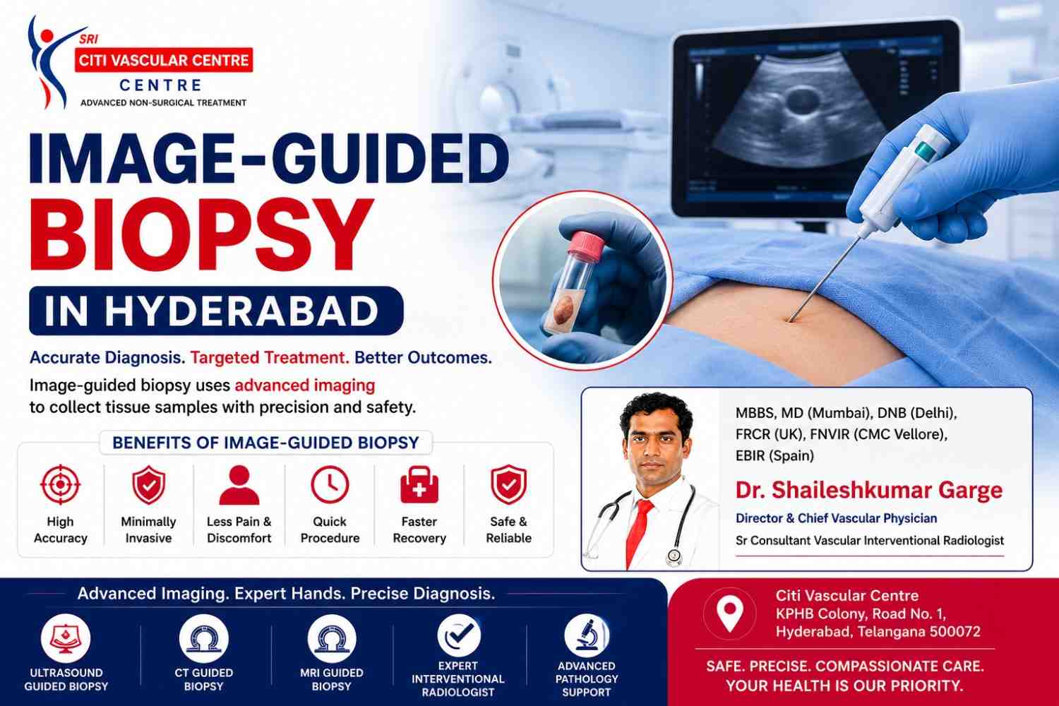
An image-guided biopsy is a minimally invasive procedure in which a small tissue sample is collected from a suspicious lump, tumour, or abnormal organ using imaging guidance such as ultrasound (USG) or CT scan. The imaging helps the doctor accurately place the biopsy needle into the target area, improving diagnostic accuracy while reducing the risk of injury to surrounding tissues. Most procedures are completed in 15–30 minutes, require only local anaesthesia, and allow patients to return home the same day.
Image-guided biopsy has become the preferred method for diagnosing many conditions because it combines precision, safety, and faster recovery. Rather than relying on surgery to obtain tissue samples, specialists can use real-time imaging to target even small or deeply located abnormalities with a tiny needle.
At Citi Vascular Centre, Hyderabad, Dr. Shaileshkumar Garge performs ultrasound-guided and CT-guided biopsies using advanced imaging equipment to obtain accurate tissue samples while maximising patient comfort and safety.
Whether a biopsy is needed to investigate a suspicious lump, confirm cancer, identify an infection, or diagnose inflammatory disease, image-guided techniques often provide reliable answers without the need for major surgery.
Book Image-Guided Biopsy — Citi Vascular Centre, KPHB, Hyderabad
Call +91-73375 83901 | WhatsApp | citivascularcentre.com | Dr. Garge FRCR (UK) | Mon–Sat 9AM–6PM
|
Feature |
Detail |
|
Procedure Type |
Core Needle Biopsy (CNB) or Vacuum-Assisted Biopsy (VAB) — under real-time image guidance |
|
What Is Collected |
A tissue core (cylinder) — provides histology and full tissue architecture, not just loose cells |
|
Imaging Used |
Real-time ultrasound (USG) | CT-guided | MRI-guided (selected centres and sites) |
|
Needle Size |
14–20 gauge spring-loaded or vacuum-assisted cutting needle |
|
Anaesthesia |
Local anaesthesia always used — skin and needle tract numbed before biopsy begins. Sedation available for deep or painful sites. |
|
Procedure Duration |
20–30 min superficial | 30–45 min deep organs. Total visit including observation: 3–6 hours. |
|
Hospital Stay |
Day-care — 2–6 hours observation post-procedure. No overnight admission for most patients. |
|
Result Timeline |
Histopathology: 3–7 working days | Immunohistochemistry (IHC) stains: up to 10–14 days |
|
Pain Level |
Mild to moderate during procedure — fully controlled with local anaesthesia. Aching 4–12 hours post-procedure. |
|
Organs Biopsied |
Liver | Kidney | Lung | Breast | Lymph node | Bone | Soft tissue | Retroperitoneum | Adrenal | Prostate |
|
Blood Tests Required |
Full blood count + coagulation screen (PT/INR, APTT, platelet count) — for all deep organ biopsies |
|
Blood Thinners |
Must be paused before biopsy — specific timings per drug advised at booking |
|
Key Advantage Over FNAC |
Tissue architecture + IHC — essential for lymphoma sub-typing, prostate Gleason grading, fibrosis staging, sarcoma classification, molecular profiling |
An image-guided biopsy is a diagnostic procedure in which a doctor uses medical imaging to guide a biopsy needle precisely into an abnormal area and obtain a tissue sample for laboratory examination.
Unlike traditional surgical biopsies, image-guided biopsies require only a small needle puncture rather than a large incision. Real-time imaging enables the doctor to visualise the needle throughout the procedure, improving accuracy and reducing the likelihood of complications.
The collected tissue is then examined by a pathologist under a microscope to determine whether the abnormality is:
Benign (non-cancerous)
Malignant (cancerous)
Inflammatory
Infectious
Autoimmune
Fibrotic or scar tissue
Another specific disease process
Because the procedure is minimally invasive, image-guided biopsy has become the standard approach for diagnosing many abnormalities in the breast, thyroid, liver, kidneys, lungs, lymph nodes, soft tissues, and other organs.
Why Is Image-Guided Biopsy Performed?
Doctors recommend an image-guided biopsy when imaging tests identify an abnormal area that requires further evaluation.
The procedure helps answer important diagnostic questions, including:
Is the lesion cancerous or non-cancerous?
What type of tumour is present?
Is there evidence of infection?
Is inflammation causing the abnormality?
Has a known cancer spread to another organ?
What is the best treatment approach?
Obtaining a tissue diagnosis often helps avoid unnecessary surgery and enables doctors to plan the most appropriate treatment based on accurate laboratory findings.
|
Type |
How It Works |
Best Used For |
Notes |
|
Core Needle Biopsy (CNB) |
Spring-loaded cutting needle fires through lesion in 0.1 seconds |
Liver, kidney, soft tissue, lymph node, thyroid (selected), peripheral lung |
Most common type. 14–18G. 2–4 cores per session. |
|
Vacuum-Assisted Biopsy (VAB) |
Rotating cutter with vacuum suction — multiple cores through one skin entry |
Breast lesions | Small deep lesions | Microcalcifications on mammogram |
Larger tissue volume. Preferred for breast and small lesions needing larger sample. |
|
Bone Trephine Biopsy |
Drill-action cutting needle through cortical bone into target |
Bone lesions, metastases, vertebral lesions, pelvic masses |
CT guidance standard. Local anaesthesia + IV sedation usual. |
|
Coaxial Technique |
Outer sheath inserted first; multiple core passes through same skin puncture |
Lung, liver, deep retroperitoneal masses |
One skin entry point for multiple samples — reduces complication risk for deep organs. |
1.Ultrasound-Guided Biopsy
Ultrasound guidance is commonly used for lesions that can be clearly visualised with ultrasound, including the thyroid, breast, liver, kidneys, lymph nodes, and superficial soft tissue masses.
Advantages include:
Real-time needle guidance
No radiation exposure
Quick procedure
Widely available
Excellent patient comfort
2.CT-Guided Biopsy
Computed tomography (CT) guidance is particularly useful for abnormalities located deep within the body, especially those that cannot be adequately visualised using ultrasound.
Common indications include:
Lung nodules
Deep abdominal masses
Retroperitoneal lesions
Pelvic tumours
Bone lesions
CT imaging allows highly accurate needle placement while avoiding important blood vessels, nerves, and other critical structures.
3.Fluoroscopy-Guided Biopsy
Fluoroscopy uses continuous X-ray imaging and is occasionally used for selected bone lesions or procedures involving contrast guidance.
Although less commonly required than ultrasound or CT guidance, it remains valuable in certain specialised situations.
4.MRI-Guided Biopsy
MRI-guided biopsy is reserved for selected lesions that are visible only on MRI, particularly some breast abnormalities.
Although highly accurate, it is generally less commonly performed than ultrasound-guided or CT-guided biopsy due to cost and availability.
|
Feature |
Ultrasound-Guided |
CT-Guided |
|
Radiation |
None |
Uses CT radiation |
|
Real-Time Imaging |
Yes |
Intermittent CT imaging |
|
Best For |
Thyroid, breast, liver, kidney, lymph nodes |
Lung, deep abdomen, pelvis, bone |
|
Procedure Time |
Usually shorter |
May be slightly longer |
|
Accessibility |
Widely available |
Ideal for deep lesions |
|
Accuracy |
Excellent |
Excellent |
Both techniques are highly effective, with the choice depending on lesion location and visibility.
Image-Guided Biopsy vs Surgical Biopsy
|
Feature |
Image-Guided Biopsy |
Surgical Biopsy |
|
Incision |
Tiny needle puncture |
Surgical incision |
|
Anaesthesia |
Usually local |
Often general or regional |
|
Hospital Stay |
Same day |
May require admission |
|
Recovery |
Quick |
Longer |
|
Scarring |
Minimal |
More noticeable |
|
Cost |
Generally lower |
Usually higher |
|
Return to Routine |
Faster |
Slower |
Both procedures are minimally invasive and image-guided — but they serve different diagnostic purposes. FNAC is quicker, less invasive, and usually sufficient for screening common lesions at thyroid, breast, and lymph node sites. Biopsy is the right choice when tissue architecture is needed, when FNAC has returned an indeterminate result, or when specific diagnosis — such as lymphoma sub-type or sarcoma classification — will directly determine the treatment regimen.
|
Parameter |
FNAC |
Core Needle Biopsy |
|
What Is Collected |
Individual cells — cytology |
Tissue cylinder — histology with architecture |
|
Needle Size |
21–25G fine aspiration needle |
14–18G spring-loaded cutting needle |
|
Anaesthesia |
Often not needed |
Always — local anaesthetic to skin and tract |
|
Duration |
10–20 minutes |
20–45 minutes |
|
Result Time |
2–5 days (cytology) |
3–7 days histology | Up to 14 days with IHC |
|
Can Sub-Type Lymphoma? |
No — FNAC insufficient for reliable lymphoma sub-typing |
Yes — tissue architecture + IHC panel provides full classification |
|
Prostate Cancer Grading |
Not appropriate for prostate |
Yes — Gleason/ISUP grade from tissue cores |
|
Molecular Profiling (EGFR, PD-L1) |
Limited by small cell numbers |
Yes — adequate tissue for IHC, FISH, and molecular markers |
|
When Preferred |
First-line for thyroid, breast, lymph node | Quick initial screening |
After inconclusive FNAC | Lymphoma | Prostate | Bone | Sarcoma | Molecular profiling needed |
Dr. Garge advises which procedure is right for your specific lesion after reviewing your imaging. Many patients can start with FNAC. Others — particularly those with suspected lymphoma, prostate lesions, or after an inconclusive FNAC — benefit from proceeding directly to core needle biopsy. The decision is always anatomy- and indication-specific.
|
Clinical Situation |
Why Biopsy Is the Right Test |
|
FNAC returned inconclusive, suspicious, or inadequate result |
Tissue architecture usually resolves indeterminate cytology. Core biopsy is the natural next step after inconclusive FNAC. |
|
Lymphoma is suspected |
Lymphoma sub-typing — DLBCL, Hodgkin, follicular, mantle cell — is essential for selecting the right chemotherapy. Cytology alone cannot reliably achieve this. |
|
Liver mass requiring definitive diagnosis |
HCC, cholangiocarcinoma, metastatic adenocarcinoma — histology with IHC identifies the specific tumour type and primary site of metastasis |
|
Lung nodule — cancer type and molecular markers needed |
Non-small cell vs small cell | Squamous vs adenocarcinoma | EGFR, ALK, PD-L1 status — all needed for targeted therapy selection and immunotherapy planning |
|
Kidney mass with uncertain features on CT |
Renal cell carcinoma vs oncocytoma vs angiomyolipoma — confirmed benign histology can avoid surgery entirely for some patients |
|
Prostate — MRI shows PI-RADS 4–5 lesion |
MRI-targeted biopsy achieves Gleason/ISUP grade — determines active surveillance vs surgery vs radiotherapy |
|
Bone lesion of uncertain nature |
Primary bone tumour vs metastasis vs infection — each requires completely different treatment. CT-guided trephine biopsy provides the definitive answer. |
|
Soft tissue mass — sarcoma suspected |
Sarcoma sub-typing (liposarcoma, leiomyosarcoma, synovial sarcoma) requires histology and IHC. The wrong surgical approach for the wrong sarcoma sub-type results in inadequate margins. |
|
Organ |
Guidance Used |
Common Indications |
Clinical Notes |
|
Liver |
USG (preferred) |
Hepatic mass, HCC, metastases, liver fibrosis staging, unexplained liver disease |
Coaxial technique standard. Post-biopsy scan confirms no haematoma. |
|
Kidney |
USG or CT |
Renal mass — RCC, oncocytoma, TCC; native kidney disease; transplant kidney |
Avoids surgery for lesions confirmed benign or metastatic on histology. |
|
Lung |
CT-guided |
Peripheral and central lung nodule, lung mass, pleural-based lesion |
Post-procedure chest X-ray standard. Pneumothorax risk 1–15%. Coaxial preferred. |
|
Breast |
USG (preferred) or stereotactic |
Breast mass, BI-RADS 4–5 lesion, microcalcifications on mammogram |
Vacuum-assisted biopsy for microcalcifications. Post-biopsy clip placed to mark site. |
|
Lymph Node |
USG |
Lymphoma sub-typing, metastatic cancer, TB with FNAC inconclusive |
Core biopsy provides adequate tissue for IHC — essential for lymphoma sub-typing. |
|
Bone |
CT-guided |
Bone metastasis, primary bone tumour, vertebral lesion, pelvic mass |
Trephine needle drills through cortex. Local anaesthesia + IV sedation for spine. |
|
Retroperitoneum |
CT-guided |
Retroperitoneal mass, para-aortic nodes, adrenal lesion |
CT essential for deep retroperitoneal structures not safely accessible by ultrasound. |
|
Soft Tissue |
USG |
Subcutaneous and intramuscular masses, suspected sarcoma |
Biopsy tract planned along future surgical excision axis — en-bloc removal if sarcoma confirmed. |
The imaging technique selected depends on which method provides the safest and most direct access to the lesion while minimising risk to surrounding structures.
Performing a biopsy without image guidance is like navigating a city without a map — it might work for large, obvious landmarks, but it becomes unreliable and potentially dangerous the moment you need precision. For a cutting needle that must travel through healthy liver parenchyma, avoid a portal vein branch, and arrive precisely at the edge of a 2cm hepatic lesion — real-time imaging is not a luxury, it is a clinical requirement.
|
Parameter |
Without Image Guidance |
With Real-Time Image Guidance |
|
Needle placement |
Estimated — high miss rate for deep lesions |
Confirmed on screen at every millimetre |
|
Targeting viable tumour vs necrosis |
Cannot distinguish — samples whatever is hit |
Solid, viable, enhancing component selected — necrotic centre avoided |
|
Vessel avoidance |
Cannot see vessels — haemorrhage risk |
Doppler (USG) or contrast CT maps vessels before needle is placed |
|
Depth measurement |
Estimated — overshoot and undershoot risk |
Precise skin-to-lesion depth measured — exact needle advance confirmed |
|
Post-biopsy assessment |
Cannot detect complications immediately |
Post-biopsy scan confirms no haematoma or pneumothorax before patient leaves recovery |
At Citi Vascular Centre, KPHB, Dr. Garge operates the ultrasound himself throughout every USG-guided biopsy — it is not delegated to a technician. For CT-guided procedures, he reviews the pre-procedure CT, plans the biopsy approach, performs the targeting CT, and advances the needle under CT fluoroscopy. The same specialist plans and executes — there is no handover between imaging and the proceduralist.
Most patients describe the procedure as causing only mild discomfort.
Local anaesthesia numbs the skin and deeper tissues before the biopsy needle is introduced. Patients may feel slight pressure or brief discomfort during tissue sampling, but significant pain is uncommon.
After the biopsy, mild soreness at the puncture site usually resolves within a day or two.
Which Anaesthesia Is Used?
|
Procedure - |
Anaesthesia |
|
Most USG-guided biopsies - |
Local anaesthesia |
|
CT-guided biopsy - |
Local anaesthesia |
|
Children - |
Sedation if required |
|
Complex procedures - |
Sedation in selected cases |
General anaesthesia is rarely required.
Preparation for image-guided biopsy is more involved than for FNAC — primarily because the larger cutting needle creates a small but real risk of bleeding that must be assessed and managed before the procedure. Dr. Garge's team will provide complete pre-procedure instructions when your appointment is booked.
|
When |
Preparation Step |
Notes |
|
At Booking |
Gather ALL imaging — CT, MRI, PET, ultrasound reports and discs |
Essential for approach planning and confirming the correct target |
|
5–7 Days Before |
Stop blood-thinning medications |
Aspirin: 5–7 days | Clopidogrel: 5–7 days | Warfarin: 5 days (INR checked Day 0) | Apixaban, Rivaroxaban: 48–72 hours. Never stop without medical guidance. |
|
2–3 Days Before |
Blood tests: full blood count, PT/INR, APTT, platelet count |
Required for all deep organ biopsies. Superficial biopsies may not need all tests. Results reviewed before confirming the procedure. |
|
Before Procedure |
Declare any bleeding disorder, liver disease, or kidney impairment |
These affect bleeding risk and may require additional precautions or dose adjustment |
|
Day of Procedure |
Fasting — 4–6 hours for deep organ biopsies |
Liver, kidney, lung, bone — 4–6 hours nil by mouth. Superficial biopsies: fasting usually not needed. Confirmed when you book. |
|
Day of Procedure |
Wear comfortable, accessible clothing |
Two-piece separates for abdominal or chest biopsy | Front-open top for breast | Loose neckwear for neck procedures |
|
Transport |
Arrange a driver home for deep organ biopsies |
IV sedation (if used) means no driving. Even without sedation, post-procedure rest is strongly advised for deep organ biopsies. |
Histopathology reports are typically available within 3 to 7 working days. When special immunohistochemistry (IHC) stains are required — for lymphoma sub-typing, identifying the primary site of a metastasis, hormone receptor status in breast cancer, or molecular markers such as EGFR, ALK, and PD-L1 in lung cancer — the full report may take up to 10 to 14 days. Dr. Garge will contact you when the complete report is ready and arrange a follow-up consultation to review the findings alongside your imaging.
|
Result Category |
What It Means |
Common Examples |
Typical Next Steps |
|
Benign |
No malignancy. Confirmed benign pathology. |
Renal oncocytoma | Hepatic haemangioma | Reactive lymph node | Lipoma |
Reassurance. Surgery avoided in many. Surveillance plan arranged. |
|
Malignant — Primary Cancer |
Cancer originating from the biopsied organ |
HCC | Renal cell carcinoma | NSCLC | Breast carcinoma |
Urgent oncology/surgery referral. Staging investigations started. MDT discussion. |
|
Malignant — Metastasis |
Secondary cancer. IHC identifies the primary site. |
Liver met from colorectal | Bone met from breast | Lymph node met from lung |
Primary tumour identified. Systemic treatment planned. Avoids exploratory surgery. |
|
Lymphoma |
Lymphoid malignancy — sub-typed by IHC and FISH |
DLBCL | Follicular lymphoma | Hodgkin lymphoma | Mantle cell lymphoma |
Sub-type determines chemotherapy regimen. Core biopsy essential — FNAC cannot sub-type lymphoma. |
|
Inflammatory or Infective |
Tissue inflammation or infection, not cancer |
TB granuloma | Sarcoidosis | Abscess | Reactive change |
Appropriate antimicrobial or anti-inflammatory treatment. Anti-TB therapy if TB confirmed. |
|
Inconclusive or Insufficient |
Core not representative of the lesion |
Necrotic tissue sampled | Heavily fibrotic lesion | Technical difficulty |
Repeat biopsy with modified approach or surgical excision biopsy. |
Immunohistochemistry (IHC): Many biopsy reports require additional IHC staining beyond the standard H&E stain — using antibody panels to identify specific proteins in the tissue. IHC determines tumour type and grade, hormone receptor status (oestrogen, progesterone, HER2 in breast cancer), PD-L1 expression for immunotherapy eligibility, EGFR and ALK mutations in lung cancer, and the primary site of a cancer that has spread. These stains add 3–7 days to reporting time but are essential for correct treatment selection.
|
Timeframe |
What to Expect |
Activity Guidance |
|
Immediately after |
2–6 hours observation. Post-biopsy scan performed. Vital signs monitored every 30–60 minutes. |
Bed rest during observation period. Arrange a driver — do not drive after deep organ biopsy. |
|
Day of Procedure |
Mild to moderate aching at biopsy site. Possible small bruise. After lung biopsy, a trace of blood in sputum is expected and normal. |
Rest at home. Paracetamol 500–1000mg as needed. Avoid strenuous activity. Light meals when tolerated. |
|
Day 1–2 |
Aching reduces progressively. Bruise may appear and fade over days. |
Light normal activities from Day 1. Office work from Day 2 for superficial biopsies. Day 3–5 for deep organ biopsies. |
|
Day 3–7 |
Awaiting histopathology result. No ongoing procedure symptoms expected. |
Normal activities fully resumed. Contact Dr. Garge team when result is ready. |
|
Follow-up |
Dr. Garge reviews result with you — correlates histopathology with imaging, explains findings, plans next steps or referral. |
Attend follow-up. Write down questions in advance. Bring all imaging. |
Call Citi Vascular Centre (+91-73375 83901) immediately after biopsy if you notice: pain worsening rather than improving after the first few hours | significant swelling or expanding bruise at biopsy site | fever above 38 degrees Celsius | shortness of breath or chest pain after upper abdominal or lung biopsy | blood in urine beyond a faint trace after kidney biopsy | shoulder tip pain after liver biopsy. These symptoms require prompt assessment.
Image-guided biopsy is generally considered a safe procedure when performed by experienced specialists using appropriate imaging guidance.
However, as with any invasive procedure, complications can occur. Your doctor will explain the specific risks before the procedure
|
Complication |
Frequency |
Severity |
Management |
|
Pain and bruising at site |
Very common |
Mild |
Paracetamol or ibuprofen. Resolves within 1–3 days. |
|
Small haematoma |
Common |
Usually minor |
Resolves spontaneously. Monitored on post-biopsy imaging. |
|
Significant bleeding |
0.5–2% deep organs |
Moderate–serious |
Angiographic embolisation in most cases. Rarely requires surgery. |
|
Pneumothorax (lung only) |
1–15% |
Variable |
Small: observation, spontaneous resolution. Large: aspiration or chest drain. |
|
Infection |
< 1% |
Moderate if untreated |
Sterile technique prevents this. Short antibiotic course if confirmed. |
|
Bile leak (liver only) |
< 0.1% |
Potentially significant |
Risk minimised by avoiding large bile ducts on imaging. Drainage if occurs. |
|
Vasovagal reaction |
Uncommon |
Momentary |
Procedure paused. Patient repositioned. Resolves promptly. |
|
Needle track seeding |
Extremely rare |
Theoretical |
Biopsy tract planned along future surgical axis for suspected sarcoma cases. |
|
Definitive tissue diagnosis |
Histology with IHC — the most complete tissue characterisation available short of open surgical biopsy. |
|
|
No general anaesthesia |
Local anaesthesia for most procedures — no intubation, no recovery room, no anaesthetic risk. |
|
|
No surgical incision |
2–3mm skin nick only. No wound, no stitches in most cases. Same-day discharge. |
|
|
Avoids unnecessary surgery |
Benign histology (renal oncocytoma, hepatic haemangioma) confirmed — operation avoided entirely. |
|
|
Enables precise treatment selection |
Lymphoma sub-type | Gleason grade | EGFR/ALK/PD-L1 status — determines exactly which therapy to use. |
|
|
Multiple sites in one session |
Liver + lymph node + peritoneal deposit biopsied in one visit — simultaneous staging and diagnosis. |
|
|
Rapid diagnosis |
3–7 days histopathology vs weeks for surgical biopsy pathway — treatment planning begins faster. |
Image-guided biopsy is appropriate for most patients with a solid lesion in an accessible organ where tissue diagnosis is required before treatment can be planned. It is particularly valuable when FNAC has been inconclusive, when lymphoma is the primary clinical suspicion, when molecular profiling for targeted therapy is needed alongside diagnosis, or when knowing whether a lesion is benign could avoid a major operation.
Biopsy requires specific precautions or may not be appropriate in certain situations. Severely deranged coagulation (INR above 1.5 or platelets below 50,000) needs correction before the procedure. Lesions with no safe needle path that avoids major vessels or critical structures may require an alternative approach. Patients who cannot remain still during the procedure — or those where surgical excision provides both diagnosis and treatment simultaneously — may be better served by a surgical approach. Dr. Garge reviews every case individually and will advise clearly at consultation.
|
Credential / Feature |
Detail |
|
Qualifications |
MBBS | MD (Mumbai) | DNB (Delhi) | FRCR (UK) | FNVIR (CMC Vellore) | EBIR (Spain/Europe) | Fellowship (North Carolina, USA) |
|
Role |
Director and Chief Vascular Physician | Senior Consultant Vascular and Interventional Radiologist |
|
Experience |
12+ years dedicated interventional radiology | 15,000+ image-guided diagnostic and interventional procedures |
|
Organs Biopsied |
Liver | Kidney | Lung (peripheral) | Breast | Lymph nodes | Bone | Soft tissue | Retroperitoneum | Adrenal | Thyroid (selected) |
|
Personal Imaging Guidance |
Dr. Garge personally operates USG throughout biopsy and plans and executes all CT-guided procedures — not split between radiologist and proceduralist |
|
One-Stop Visit |
Pre-biopsy imaging review + biopsy + immediate post-biopsy scan all in one appointment — no multi-department scheduling |
|
IHC Coordination |
Works with experienced histopathology partners for full IHC panel — lymphoma sub-typing, hormone receptors, molecular markers, primary site identification |
|
Post-Biopsy Follow-Up |
Result consultation with Dr. Garge — histopathology reviewed alongside imaging, next steps explained, oncology or surgical referral coordinated |
|
Centre |
High-resolution USG with colour Doppler | CT access | Sterile procedure room | Day-care ward | Insurance assistance | KPHB Colony, Hyderabad |
Citi Vascular Centre is a dedicated centre for minimally invasive image-guided procedures. Every biopsy is planned using modern imaging technology with emphasis on accuracy, patient safety, and timely diagnosis.
Our Advantages
Advanced Imaging
2.Minimally Invasive Care
3.Day-Care Procedures
4.Patient-Centred Care
5. Comprehensive Diagnostic Support
6.Convenient Location
|
Centre |
Contact |
Hours |
|
Citi Vascular Centre |
+91-73375 83901 |
KPHB Colony, Road No. 1, Hyderabad, Telangana 500072 | Mon–Sat 9AM–6PM |
|
Book Online |
citivascularcentre.com |
WhatsApp + online booking | Pre-biopsy imaging review included | Insurance assistance available |
Easily accessible from:
Miyapur
Hitech City
Gachibowli
Madhapur
Ameerpet
Bachupally
Nizampet
Secunderabad
|
1 |
Registration and review of all imaging — CT, MRI, PET, previous ultrasound. Blood test results confirmed adequate for biopsy. |
|
2 |
Consultation with Dr. Garge — target lesion confirmed, biopsy approach planned on imaging, consent obtained, sedation discussed if relevant. |
|
3 |
Fresh pre-biopsy imaging. USG Doppler maps vessels around the lesion. CT localising scan for CT-guided procedures. Needle path planned to avoid critical structures. |
|
4 |
Local anaesthetic administered at the skin, subcutaneous tissue, and target organ capsule. IV cannula and sedation if required. 10 minutes allowed for full anaesthetic effect. |
|
5 |
2–3mm skin nick made. Biopsy needle advanced under continuous real-time imaging to lesion. Position confirmed. Spring-loaded cutter fires — 0.1 seconds. 2–4 cores extracted from different lesion zones and placed in formalin. |
|
6 |
Immediate post-biopsy imaging scan confirms no haematoma or pneumothorax before patient is moved to recovery. Result confirmed to Dr. Garge. |
|
7 |
2–6 hours observation in recovery. Vital signs monitored. Pain assessed and managed. Oral fluids offered when appropriate. Driver arranges collection. |
|
8 |
Discharge with written post-procedure instructions, contact number for concerns, and follow-up appointment details. Histopathology report reviewed with Dr. Garge in 3–7 days. |
Important before your biopsy: (1) Bring all imaging discs and reports — not just the written report. Dr. Garge uses the actual scan images to plan the needle approach. (2) Confirm with our team which blood thinners to stop and when. (3) Arrange a driver home for any deep organ biopsy. (4) Have something to eat before the observation period ends — light meal is encouraged during recovery.
Q1: What is image-guided biopsy and how is it different from FNAC?
Image-guided biopsy uses a spring-loaded cutting needle (14–18G) under real-time ultrasound or CT to extract a tissue core for histopathological examination. FNAC uses a fine needle (21–25G) to aspirate loose cells for cytology. Biopsy provides full tissue architecture — essential for lymphoma sub-typing, prostate Gleason grading, sarcoma classification, liver fibrosis staging, and molecular profiling that cytology cannot reliably achieve.
Q2: Is image-guided biopsy painful?
More uncomfortable than FNAC, but well-controlled with local anaesthesia. The skin, subcutaneous tissue, and needle tract are numbed fully before the biopsy needle is inserted. The cutting action takes 0.1 seconds — most patients feel pressure, not pain. Post-procedure aching for 4–12 hours is typical and managed comfortably with paracetamol. IV sedation is available for deeper or more sensitive sites such as bone and retroperitoneum.
Q3: Which is the best hospital for image-guided biopsy in Hyderabad?
Citi Vascular Centre, KPHB Colony, Road No. 1, Hyderabad, led by Dr. Shaileshkumar Garge — FRCR (UK), FNVIR (CMC Vellore), EBIR (Spain) — is one of Hyderabad's most credentialled centres for USG and CT-guided biopsy. High-resolution real-time imaging, post-biopsy scan included, full IHC coordination, and a result follow-up consultation with Dr. Garge are all provided. Call +91-73375 83901.
Q4: How long does an image-guided biopsy take?
The biopsy itself takes 20–30 minutes for superficial sites (breast, lymph node, soft tissue) and 30–45 minutes for deep organs (liver, kidney, lung, bone). Total clinic time — including consultation, pre-procedure imaging, the biopsy, post-biopsy scan, and 2–6 hours observation in recovery — is typically 4–6 hours for deep organ biopsies. Arrange a driver for all deep organ procedures.
Q5: Do I need to stop blood thinners before a biopsy?
Yes — blood-thinning medications must be paused before most biopsies. Recommended pause durations: aspirin 5–7 days | clopidogrel 5–7 days | warfarin 5 days (INR checked on the day) | apixaban and rivaroxaban 48–72 hours. Specific timings depend on the organ being biopsied and your individual bleeding risk. Never stop anticoagulants without medical guidance — Dr. Garge's team will give you written instructions when you book.
Q6: When will I get my biopsy results?
Standard histopathology reports take 3–7 working days. When additional immunohistochemistry (IHC) stains are required — for lymphoma sub-typing, identifying a cancer's primary site, hormone receptor status, or molecular markers such as EGFR, ALK, and PD-L1 — the full report takes up to 10–14 days. Dr. Garge will contact you when the complete report is available and arrange a consultation to explain the findings alongside your imaging.
Q7: What is the risk of pneumothorax with a lung biopsy?
Pneumothorax — air entering the space between lung and chest wall — occurs in approximately 5–15% of CT-guided peripheral lung biopsies, varying with lesion depth and underlying lung health. Most are small and resolve without treatment. Significant pneumothorax requiring aspiration or a chest drain occurs in approximately 1–3% of cases. A chest X-ray is performed before discharge from Citi Vascular Centre to detect pneumothorax before you leave.
Q8: Can a biopsy diagnose lymphoma?
Yes — and core needle biopsy is the preferred method because lymphoma sub-typing requires tissue architecture and adequate tissue volume for a full immunohistochemistry panel. FNAC alone cannot reliably sub-type lymphoma. The specific sub-type — DLBCL, follicular, Hodgkin, mantle cell, T-cell — determines which chemotherapy regimen is used. At Citi Vascular Centre, KPHB, USG-guided lymph node core biopsy with full IHC reporting is available.
Q9: When should I choose biopsy instead of FNAC?
Choose core needle biopsy over FNAC when: lymphoma is the primary clinical suspicion | FNAC has returned inconclusive or indeterminate | diagnosis requires tissue architecture (follicular thyroid neoplasm, liver fibrosis staging, sarcoma sub-typing) | molecular profiling (EGFR, ALK, PD-L1, HER2) is needed alongside diagnosis | prostate cancer Gleason grading is required. Dr. Garge advises which test is right for your specific lesion after reviewing your imaging. Call +91-73375 83901.
|
Image-guided biopsy extracts a tissue core under real-time USG or CT — providing histology and full tissue architecture, not just loose cells |
|
Essential for lymphoma sub-typing | prostate Gleason grading | liver fibrosis staging | sarcoma classification | EGFR/ALK/PD-L1 molecular profiling |
|
Blood thinners must be stopped before biopsy — specific timing per drug. Blood tests required for deep organ procedures. Arrange a driver home. |
|
Post-biopsy imaging scan confirms no haematoma or pneumothorax before every patient leaves Citi Vascular Centre |
|
Histopathology in 3–7 days | IHC stains up to 14 days | Results reviewed with Dr. Garge at follow-up consultation |
|
Replaces open surgical biopsy for most accessible lesions — equivalent diagnostic accuracy, far less invasive |
|
Dr. Shaileshkumar Garge FRCR (UK) | Citi Vascular Centre, KPHB | +91-73375 83901 | Mon–Sat 9AM–6PM |
When a clinical team needs a definitive tissue answer before any treatment can begin, image-guided biopsy is what provides it. A cutting needle, advanced under real-time ultrasound or CT by an experienced interventional radiologist, extracts a tissue core from the target lesion in 20 to 45 minutes under local anaesthesia. No surgery, no general anaesthetic, no overnight stay. The histopathology report that follows gives not just a cancer diagnosis but the full characterisation — sub-type, grade, molecular profile — that determines which specific treatment will work for that particular patient.
At Citi Vascular Centre, KPHB Colony, Hyderabad, Dr. Shaileshkumar Garge — FRCR (UK), FNVIR (CMC Vellore), EBIR (Spain) — performs the pre-biopsy imaging review, the biopsy itself, the post-biopsy scan, and the result consultation. There is no handover between specialists. If you have been told that a tissue diagnosis is needed before your oncologist or surgeon can proceed, this is where to begin.
Book Image-Guided Biopsy — Citi Vascular Centre, KPHB, Hyderabad
Liver | Kidney | Lung | Breast | Lymph Node | Bone | Soft Tissue | Retroperitoneum
Dr. Shaileshkumar Garge | FRCR (UK) | FNVIR (CMC Vellore) | EBIR (Spain) | 12+ Years | 15,000+ Procedures
Call +91-73375 83901 | WhatsApp Now | citivascularcentre.com
USG & CT-Guided | Local Anaesthesia | Same-Day Discharge | Histopathology + IHC | Insurance Assisted | Mon–Sat 9AM–6PM