LAST MEDICALLY REVIEW
August 2026 — Dr. Shaileshkumar Garge
Citi Vascular Hospital, KPHB Colony, Road No. 1, Hyderabad, Telangana 500072
QUICK ANSWER
What Is Gasserian Ganglion Radiofrequency Ablation for Trigeminal Neuralgia?
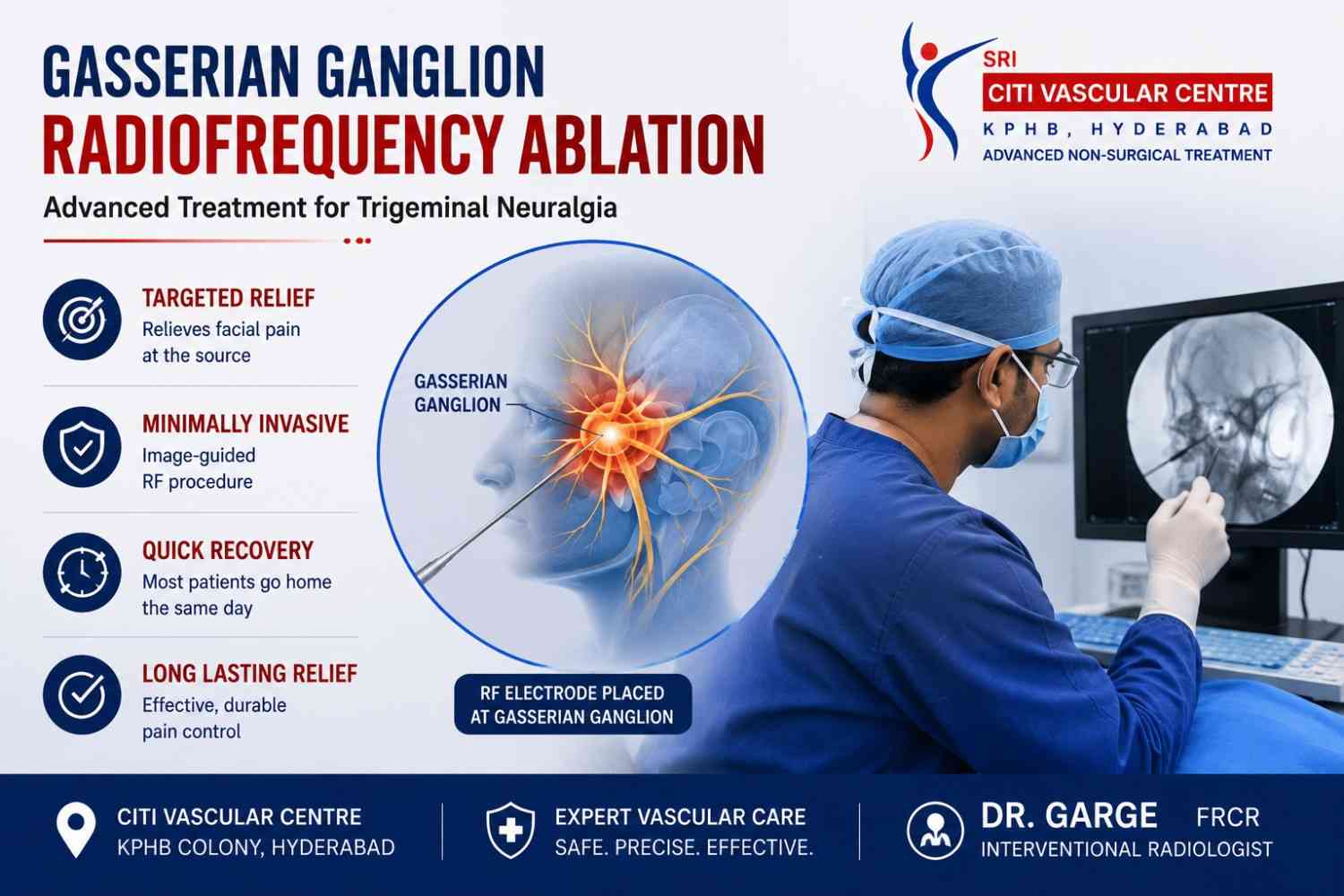
Gasserian ganglion radiofrequency ablation is a minimally invasive, fluoroscopy-guided procedure used to treat severe trigeminal neuralgia when medications fail to provide adequate or lasting relief. A thin RF electrode is positioned through the foramen ovale (a natural opening in the skull base) toward the trigeminal ganglion. Electrical stimulation identifies the painful trigeminal division (V1, V2, or V3) before controlled radiofrequency heat interrupts the abnormal pain signals. Rapid pain relief is possible, but sensory changes and recurrence should be discussed honestly before treatment. Dr. Garge FRCR (UK) | Citi Vascular Centre, KPHB, Hyderabad | +91-73375 83901.
Trigeminal neuralgia is a neurological condition characterised by sudden, severe, electric shock-like facial pain that may be triggered by activities such as eating, speaking, brushing the teeth, or touching the face.— an electric shock or lightning bolt striking the face, triggered by something as gentle as a breeze, a sip of water, or brushing a tooth. For patients whose pain has not been controlled by medication, the prospect of continuing to live in fear of the next attack — every meal, every conversation, every attempt to wash the face — is not sustainable. This is where interventional treatments like Gasserian ganglion radiofrequency ablation can offer a meaningful alternative.
This page explains what radiofrequency ablation of the Gasserian ganglion is, how it works, who is suitable, the step-by-step procedure, the risks — including the specific concerns around corneal sensation for V1 involvement — how it compares with surgery and radiosurgery, and what recovery looks like. It is written for patients who want to understand the treatment thoroughly before making a decision — not for patients who simply want to be reassured. Treatment planning involves clinical assessment, review of relevant imaging, evaluation of previous medication, and discussion of the potential benefits, risks, and alternative treatments with the patient.
Trigeminal Neuralgia Assessment — Citi Vascular Centre, KPHB, Hyderabad
Call +91-73375 83901 | WhatsApp 73375 83901 | citivascularcentre.com | Mon–Sat 9AM–6PM
Book your Appointment here: | Appoinment: |Call +91-7032508468| Whatsapp:+91-7337583901
|
Feature |
Detail |
|
Procedure Type |
Minimally invasive percutaneous neuroablative procedure — no open surgery, no craniotomy |
|
Guidance |
Fluoroscopy throughout — skull base anatomy, foramen ovale access, and electrode positioning |
|
Energy Used |
Radiofrequency electrical energy — generates controlled heat at the electrode tip to create a precise lesion in the trigeminal ganglion |
|
Target |
Gasserian (trigeminal/semilunar) ganglion — the cluster of sensory nerve cell bodies giving rise to V1, V2, and V3 |
|
Access Route |
Foramen ovale — a natural opening in the base of the skull through which the mandibular (V3) division passes |
|
Division Targeting |
Sensory stimulation identifies V1 (forehead/eye), V2 (cheek/upper lip), or V3 (jaw/chin) before energy delivery |
|
Anaesthesia |
Local anaesthesia + IV sedation — patient must be able to communicate stimulation responses; excessive sedation interferes with targeting |
|
Hospital Stay |
Hospital stay: Depending on the patient's clinical condition, the procedure and recovery, discharge may be possible on the same day or after overnight observation. |
|
Pain Relief |
Pain relief: Improvement may occur within hours to days after treatment, although the response and duration of relief vary between patients. |
|
Sensory Trade-off |
Some degree of facial numbness or altered sensation is common — an expected consequence of the neuroablative mechanism |
Trigeminal neuralgia (TN) is a neurological pain disorder affecting the trigeminal nerve — the major sensory nerve of the face. Trigeminal neuralgia causes sudden, severe, electric shock-like facial pain that may last from a fraction of a second to approximately two minutes per attack. Common triggers include eating, speaking, brushing the teeth and touching the face. Eating, drinking, speaking, washing the face, and even cool air can trigger an attack. The attacks themselves last seconds to minutes — but their unpredictability and severity, and the fear they generate between attacks, make trigeminal neuralgia a condition that profoundly affects quality of life.
|
Division |
Name |
Facial Area |
Typical Pain Location |
|
V1 |
Ophthalmic |
Forehead, eye, upper nose |
Forehead, brow, upper eyelid, nasal bridge |
|
V2 |
Maxillary |
Cheek, upper lip, upper teeth |
Cheek, nasolabial area, upper lip, upper gum, side of nose |
|
V3 |
Mandibular |
Lower jaw, chin, lower teeth |
Lower jaw, chin, lower lip, lower teeth, inner cheek |
The most common pattern in classical trigeminal neuralgia involves V2 or V3, or both — but V1 involvement occurs in approximately 5% of cases and requires additional caution during any interventional treatment because of the cornea's dependence on ophthalmic division sensory innervation. For patients whose pain has not been adequately controlled by carbamazepine, oxcarbazepine, or other medications — or who cannot tolerate the medication side effects — interventional treatments including Gasserian ganglion RF ablation become important options to discuss.
The Gasserian ganglion — also called the trigeminal ganglion or semilunar ganglion — is a collection of sensory nerve cell bodies associated with the trigeminal nerve. It is located inside a small anatomical space called Meckel's cave, near the base of the skull. All three trigeminal divisions (V1, V2, and V3) arise from this structure, arranged in a predictable spatial relationship that The trigeminal ganglion is an anatomical target for selected percutaneous procedures used to manage trigeminal neuralgia. The choice of target and treatment depends on the patient's clinical findings and the procedure being considered.Because the three divisions are anatomically organised in relation to the ganglion, a carefully positioned RF electrode — confirmed by electrical stimulation — can selectively treat the division that is causing the patient's pain rather than destroying the entire nerve.
The foramen ovale — a natural opening in the greater wing of the sphenoid bone at the skull base — provides the percutaneous access route to the Gasserian ganglion. The mandibular division (V3) passes through this opening, and the cannula used in Gasserian ganglion procedures is directed through this foramen under fluoroscopic guidance. The foramen ovale approach avoids the need for surgical craniotomy and allows the procedure to be performed under local anaesthesia and sedation rather than full general anaesthesia.
Gasserian ganglion radiofrequency ablation is a percutaneous neuroablative procedure — meaning the treatment is delivered through the skin with a needle (percutaneous) and its mechanism is the deliberate, controlled interruption of nerve function (neuroablative). The RF generator produces alternating electrical current at radiofrequency, which generates ionic friction and heat at the electrode tip. This controlled thermal lesion — typically at temperatures of 65–90°C, maintained for defined periods — creates a localised area of nerve injury that reduces the abnormal pain signal transmission responsible for trigeminal neuralgia.
It is important that patients understand the neuroablative nature of this treatment from the outset. RF ablation does not remove the cause of trigeminal neuralgia (neurovascular compression, for example). It interrupts the pain signals by deliberately injuring selected nerve fibres. Because the trigeminal nerve also carries normal facial sensation, some degree of numbness or altered sensation in the treated distribution is a predictable consequence — not a complication in the unexpected-event sense, but an expected trade-off of the neuroablative mechanism. The goal is to achieve meaningful pain control while minimising unnecessary sensory disruption — and this requires careful targeting through electrical stimulation.
Book your Appointment here: | Appoinment: |Call +91-7032508468| Whatsapp:+91-7337583901
RF May Be Appropriate When:
Confirmed trigeminal neuralgia diagnosis | Medication has failed or caused unacceptable side effects | Patient is not suitable for open neurosurgery (MVD) due to age or comorbidity | MRI does not show convincing neurovascular compression that MVD could address | Patient prefers a minimally invasive approach | Rapid pain relief is a priority | Previous treatment has been successful but pain has recurred
Consider Alternatives When:
MRI findings suggestive of clinically significant neurovascular compression may support consideration of microvascular decompression in appropriately selected patients. The potential benefits, risks and limitations should be discussed before treatment. | Patient is a good surgical candidate who prefers no facial numbness | V1 is the primary involvement and corneal risk requires careful counselling | Diagnosis is uncertain | Patient cannot cooperate with stimulation responses during procedure
Important: There is no single treatment that is suitable for every patient with trigeminal neuralgia. Treatment options may include medication, microvascular decompression, radiofrequency procedures and stereotactic radiosurgery. The choice depends on the diagnosis, imaging findings, previous treatment, general health and patient preferences. Each option should be discussed with its potential benefits, risks and limitations.For classical TN with convincing neurovascular compression on MRI, microvascular decompression (MVD) addresses the underlying cause without deliberately damaging the nerve — and is generally the preferred surgical option for suitable candidates. RF is most valuable when the patient is not a good MVD candidate, when MVD is declined or has previously been performed, or when a minimally invasive approach is clinically appropriate. The decision should always be made after individual assessment and honest discussion of all available options.
The following section describes the usual stages of Gasserian ganglion radiofrequency ablation, from pre-procedure assessment to discharge. Individual steps may vary according to the patient's clinical condition, anatomy and procedural requirements.Individual steps may be modified based on the patient's specific pain pattern, anatomy, and clinical requirements.
1.Pre-Procedure Assessment and MRI Review
The treating practitioner reviews the clinical history, including the affected trigeminal division, pain triggers, medications tried and baseline facial sensation. MRI findings are assessed for neurovascular compression and other possible structural causes of facial pain. — specifically which trigeminal division is involved, what triggers attacks, what medications have been tried, and the patient's baseline facial sensation including corneal reflex when V1 may be involved. MRI of the trigeminal nerve is reviewed — not only for neurovascular compression but for any other structural cause. Importantly, a blood vessel touching the trigeminal nerve on MRI is not automatically evidence of clinically significant compression — nerve displacement, indentation, or atrophy provides stronger evidence of a causative relationship.
2. Patient Positioning and Preparation
The patient is positioned on the fluoroscopy table, usually lying flat on their back. A bite block is placed between the teeth if appropriate. IV access is established. The face and submental area (below the chin) are cleaned with antiseptic. The skin entry point — typically in the cheek or submental area — is marked based on fluoroscopic planning.
3. Local Anaesthesia and Sedation
Local anaesthetic is infiltrated at the skin entry point. IV sedation is administered to make the initial needle placement comfortable while keeping the patient conscious enough to respond to stimulation. This balance — adequate comfort without excessive sedation — is one of the most important anaesthetic considerations for this procedure. A patient who is too deeply sedated cannot reliably report stimulation responses, which impairs targeting accuracy.
4. Fluoroscopy-Guided Cannula Placement
A specialised RF cannula (a hollow needle) is advanced through the skin entry point and directed toward the foramen ovale under continuous fluoroscopic guidance. The skull base anatomy — the foramen ovale, the petrous apex, and the relationship to the carotid canal — is monitored throughout. The foramen ovale is typically identified in a specific oblique fluoroscopic projection. Correct passage through the foramen ovale is confirmed before the cannula is advanced toward the Gasserian ganglion.
5. Sensory and Motor Stimulation — The Critical Targeting Step
Once the cannula tip is in the estimated position of the trigeminal ganglion, the RF electrode delivers a low-voltage electrical stimulus. The patient is asked: 'Where do you feel this sensation?' The response confirms which trigeminal division is being stimulated. V2 stimulation produces tingling in the cheek, nasolabial area, upper lip, or upper teeth. V1 stimulation produces sensation in the forehead, brow, or eye region. V3 produces sensation in the lower jaw, chin, or lower teeth. Motor stimulation at low frequency identifies the mandibular component — the masseter muscle contraction indicates proximity to V3 motor fibres. The goal is a concordant response: stimulation sensation in the same area as the patient's pain.
6. Radiofrequency Energy Delivery
Once concordant stimulation has been confirmed, the RF generator delivers controlled heat at the electrode tip. The temperature, duration, and number of lesions are individualised — higher temperatures produce larger lesions but also increase the risk of sensory complications. Published evidence suggests that outcomes do not simply improve linearly with higher temperature, and that the degree of sensory change correlates with complication risk. Many protocols use multiple lesion cycles at 65–80°C, reassessing sensation and pain response between cycles, rather than a single maximum-temperature burn.
7. Post-Procedure Assessment and Discharge
Immediately after the procedure, the treating team assesses facial sensation, pain status, jaw strength where relevant and corneal sensation or reflex when the ophthalmic division is involved. Discharge timing depends on clinical stability and the need for observation.
Gasserian ganglion RF is not simply a 'pain-blocking injection.' It is a neuroablative procedure that deliberately injures nerve tissue to reduce pain transmission. Understanding the risks — honestly and completely — is essential before making a decision about treatment.
|
Complication |
Frequency |
Clinical Notes |
|
Facial numbness and altered sensation |
Common — expected outcome |
The intended result of the neuroablative procedure — the degree varies with lesion parameters and target. Many patients accept moderate numbness in exchange for pain relief. |
|
Dysesthesia — unpleasant altered sensation |
Uncommon to common |
Tingling, burning, or uncomfortable sensation in the treated area — distinct from the original trigeminal neuralgia pain. Can be persistent and distressing in some patients. |
|
Masticatory (chewing) weakness |
Uncommon — V3-related |
Motor fibres of V3 supply the muscles of mastication. Injury to motor fibres produces jaw weakness, usually temporary, occasionally persistent. |
|
Reduced corneal sensation |
Uncommon — V1-related |
The most serious sensory complication for V1 procedures. The cornea depends on trigeminal V1 innervation for its protective blink reflex. Reduced corneal sensation can lead to corneal dryness, exposure, and ulceration if not monitored. |
|
Anesthesia dolorosa |
Rare but serious |
A numb area that develops persistent burning or painful sensations — considered one of the most feared complications of destructive trigeminal procedures. Reported frequency varies substantially between series. |
|
Vascular injury / haematoma |
Rare |
The cannula passes near important vascular structures at the skull base. Vascular injury, though uncommon, can have serious consequences. Experienced operator and fluoroscopic guidance reduce this risk. |
|
Infection / CSF leak |
Rare — < 1% |
Infection or cerebrospinal fluid leakage: These are potential complications. Their reported frequency varies between studies and clinical settings. Include a numerical estimate only when supported by an appropriate, identifiable clinical reference. |
|
Recurrence of trigeminal neuralgia pain |
Common over time — not a complication per se |
The trigeminal nerve may recover after RF, and pain can return months to years after treatment. Recurrence is a predictable long-term limitation of all neuroablative procedures. |
Corneal risk in ophthalmic division (V1) treatment: Reduced corneal sensation may impair the protective blink reflex and increase the risk of dryness, exposure and corneal injury. Patients should receive appropriate counselling and instructions to seek prompt medical attention for eye redness, pain, irritation or changes in vision.
|
Feature |
Gasserian Ganglion RF |
Microvascular Decompression (MVD) |
|
Mechanism |
Controlled heat interrupts pain-transmitting nerve fibres — neuroablative |
Separates offending blood vessel from trigeminal nerve — does not damage the nerve |
|
Surgical access |
Percutaneous — no craniotomy |
Open craniotomy required |
|
Suitable when |
No convincing neurovascular compression | Unfit for open surgery | Rapid relief priority | MVD declined or failed |
Classical TN with convincing neurovascular compression | Good surgical candidate | Preference for no facial numbness |
|
Facial numbness |
Common — expected consequence of neuroablation |
Less common — nerve is not deliberately injured |
|
Pain recurrence |
Common over years — neuroablative effect wears off as nerve recovers |
Pain recurrence after microvascular decompression varies according to patient selection, clinical findings, surgical outcomes and follow-up duration. Long-term results should be interpreted using relevant clinical evidence. |
|
Can be repeated |
Yes — repeat percutaneous treatment often possible |
Reoperation is possible but more complex than primary MVD |
Balloon compression and glycerol rhizotomy are two other percutaneous alternatives to RF — both approach the trigeminal ganglion through the foramen ovale. Balloon compression uses mechanical compression rather than heat. Glycerol rhizotomy injects a neurolytic agent. Both can provide significant pain relief; comparative studies show different complication profiles between techniques. Gamma Knife stereotactic radiosurgery uses focused radiation targeted at the trigeminal nerve root — the main practical difference from RF is the delayed onset of effect (weeks rather than hours to days), Stereotactic radiosurgery generally has a delayed onset of pain relief compared with percutaneous radiofrequency procedures. The suitability of each approach depends on the patient's clinical condition and the urgency of symptom control.
|
Timeframe |
What to Expect |
|
Immediately after procedure |
Facial sensation assessment, pain assessment, corneal reflex check (V1), jaw strength check (V3). Mild facial soreness at needle entry site. Numbness in the treated division — expected and immediate. |
|
Hours to Days 1–3 |
Most patients experience substantial relief from the electric shock-like attacks within hours to days of successful treatment. The treated area will feel numb or different — this persists and may be permanent. Mild headache and facial soreness settle within 2–3 days. |
|
Week 1–2 |
Most patients return to eating, speaking, and daily activities within 1–2 weeks. Temporary chewing weakness (V3 motor involvement) resolves over weeks in most cases. Baseline review of corneal sensation and jaw function. |
|
Month 1–3 |
Follow-up clinical assessment of pain status, facial sensation quality, any dysesthesia, corneal health (V1), and jaw strength. Assessment of whether electric shock attacks have returned and whether the treated area feels acceptable to the patient. |
|
Long-term |
Annual or as-needed review. Pain may recur months to years after treatment as the nerve recovers. Early recurrence after a brief symptom-free period should prompt reassessment — including MRI if any atypical features. |
The duration of pain relief after Gasserian ganglion radiofrequency ablation varies between patients. Published studies report different rates of initial pain relief and recurrence depending on patient selection, treatment technique, outcome definitions and follow-up duration. Any numerical estimates should be accompanied by references to the relevant studies. Patients should understand that pain may recur and that long-term relief is not guaranteed..
Gasserian ganglion radiofrequency ablation may provide prolonged pain relief, but it should not be presented as a guaranteed permanent cure. Some patients remain symptom-free for extended periods, while others experience recurrence. If pain returns, reassessment can help determine whether medication adjustment, repeat treatment or an alternative approach is appropriate.
Q1: What is Gasserian ganglion radiofrequency ablation?
A: Gasserian ganglion radiofrequency ablation is a minimally invasive, fluoroscopy-guided procedure for trigeminal neuralgia that does not respond adequately to medication. A thin RF electrode is placed through the foramen ovale (a natural skull base opening) to the trigeminal ganglion. Electrical stimulation identifies the affected facial division before controlled radiofrequency heat interrupts the pain-transmitting fibres. No open surgery. No craniotomy. Rapid pain relief is possible, though facial numbness and potential recurrence should be understood before treatment.
Q2: Is radiofrequency ablation effective for trigeminal neuralgia?
A: Radiofrequency ablation can provide pain relief in selected patients with trigeminal neuralgia. Reported outcomes vary across studies, and pain may recur during follow-up. Facial numbness, altered sensation and other complications are possible. Treatment decisions should be based on individual clinical assessment and discussion of the available alternatives.
Q3: Will I have facial numbness after the procedure?
A: Some degree of facial numbness or altered sensation in the treated trigeminal division is a common and expected outcome of Gasserian ganglion RF — because the procedure intentionally disrupts nerve fibres to reduce pain transmission. The degree of numbness varies. Some patients find it mild and acceptable. Others find persistent numbness more bothersome than anticipated. This treatment trade-off — pain relief in exchange for altered facial sensation — must be discussed honestly before the procedure. Anesthesia dolorosa (a painful numb face) is an uncommon but serious possibility.
Q4: Can RF ablation treat V2 trigeminal neuralgia?
A: Yes — the maxillary division (V2), which covers the cheek, nasolabial area, upper lip, upper teeth, and side of the nose, is one of the most common targets for Gasserian ganglion RF. Sensory stimulation during the procedure identifies V2 activation specifically — the patient reports tingling in the cheek or upper lip area — providing physiological confirmation that the electrode is targeting the right division before any radiofrequency energy is delivered.
Q5: Is V1 trigeminal neuralgia safe to treat with RF?
A: RF can be used for V1 (ophthalmic division) trigeminal neuralgia, but it requires significantly more caution than V2 or V3 treatment. The cornea receives its sensory innervation from V1 — and reduced corneal sensation from the RF lesion can impair the normal protective blink reflex, potentially leading to corneal dryness, exposure, ulceration, and permanent vision complications. V1 RF treatment requires specific pre-procedure ophthalmological assessment, very careful patient education, and prompt specialist attention if any eye symptoms develop after the procedure.
Q6: How long does the procedure take?
A: The total time at the treatment facility depends on preparation, anaesthesia, the procedure and post-procedure observation. Recovery requirements differ between percutaneous radiofrequency ablation and surgical procedures such as microvascular decompression.
Q7: Is RF ablation better than microvascular decompression for trigeminal neuralgia?
A: Neither treatment is universally better for every patient. Microvascular decompression may be considered in selected patients with classical trigeminal neuralgia and clinically significant neurovascular compression. Radiofrequency ablation may be an option for selected patients based on their clinical findings, surgical suitability and treatment preferences. The potential benefits, risks and limitations of each option should be discussed with the treating practitioner.
Q8: Can trigeminal neuralgia return after RF?
A: Yes — recurrence is one of the important long-term limitations of all neuroablative procedures including RF. Published series report that recurrence rates increase substantially with longer follow-up — many series report 25–50%+ recurrence at 5 years. The trigeminal nerve can recover after the RF lesion, and pain signals may return. If pain recurs, the patient should be reassessed — not automatically assumed to need immediate repeat treatment — as medication adjustment or an alternative procedure may also be appropriate depending on the clinical picture.
Q9: What is anesthesia dolorosa and how common is it?
A: Anesthesia dolorosa is a rare but serious complication of destructive trigeminal procedures — including RF. It describes a situation where an area of the face becomes numb (anesthesia) but simultaneously develops a persistent, burning, or deeply unpleasant pain sensation (dolorosa). It is distinct from the original trigeminal neuralgia pain and can be very difficult to treat. Its reported frequency varies significantly between published series. It is one reason that RF lesion parameters should be carefully individualised rather than using maximum-temperature ablation.
Q10: What should I watch for after Gasserian ganglion RF?
A: Contact the treating team promptly for: any vision change or blurred vision | eye redness or persistent irritation | difficulty closing the eye | severe or worsening eye pain | new facial weakness | rapidly increasing swelling | fever | severe headache | new neurological symptoms. Eye symptoms are particularly critical after V1-involved procedures. Many of these warrant same-day or emergency assessment rather than waiting for a scheduled follow-up appointment.
Q11: Who performs Gasserian ganglion RF in Hyderabad?
A: Patients seeking an assessment for trigeminal neuralgia in Hyderabad can consult an appropriately qualified medical practitioner. Evaluation may include clinical examination, review of imaging and discussion of medication, radiofrequency procedures, microvascular decompression and other appropriate treatment options.
Q12: Does a blood vessel touching the trigeminal nerve on MRI mean I need surgery?
A: Not automatically. Vascular contact with the trigeminal nerve on MRI is common and does not by itself confirm clinically significant neurovascular compression. Evidence of nerve displacement, indentation, or atrophy alongside a matching classical TN clinical pattern provides stronger support for compression. An isolated blood vessel 'touching' the nerve without these features may not be the cause of the patient's symptoms. MRI findings must always be interpreted alongside the clinical history — not used in isolation to recommend or exclude surgery.
|
Reference |
Key Finding / Relevance |
|
Bendtsen L, Zakrzewska JM et al. European Journal of Neurology. 2019. |
European Academy of Neurology guideline on trigeminal neuralgia. Establishes diagnostic criteria, medication hierarchy, and surgical/percutaneous treatment indications. Supports individualised treatment selection. |
|
Therapeutic efficacy and safety of RF ablation for TN — systematic review and meta-analysis. |
Reviewed different RF techniques, temperatures, and approaches — found high initial efficacy with variable long-term durability. Supports careful lesion parameter individualisation. |
|
Comparison of RF thermocoagulation with balloon compression — systematic review and meta-analysis. |
Comparative assessment of efficacy and adverse effects between percutaneous techniques. Informs patient counselling on complication trade-offs between RF and balloon compression. |
|
Wang JY et al. Radiofrequency thermocoagulation for trigeminal neuralgia — review. |
Summarises efficacy, technique considerations, and complications associated with RF thermocoagulation across published series. Relevant to post-procedure counselling. |
KEY POINTS
Citi Vascular Centre, KPHB Colony, Road No. 1, Hyderabad — trigeminal neuralgia RF assessment and treatment for patients from:
Citi Vascular Centre, KPHB Colony, Road No. 1, Hyderabad, Telangana 500072.
Hitech City, Ameerpet and Madhapur — 20 min | Gachibowli and Banjara Hills — 25 min
Secunderabad and Begumpet — 25 min | Telangana & AP — outstation welcome
Gasserian ganglion radiofrequency ablation is a minimally invasive treatment option for selected patients with trigeminal neuralgia that has not responded adequately to medication. The procedure uses imaging guidance to position an RF electrode near the trigeminal ganglion and deliver controlled energy to interrupt pain transmission. Outcomes vary, and treatment suitability should be determined after individual clinical assessment.
Potential benefits and limitations of Gasserian ganglion radiofrequency ablation should be discussed before treatment. Facial numbness and altered sensation may occur, ophthalmic division involvement carries specific corneal risks, and pain may recur over time. Microvascular decompression and other treatment options may be appropriate for selected patients. The treatment decision should consider the clinical diagnosis, expected benefits, risks, patient preferences and quality of life.
Trigeminal Neuralgia RF Treatment — Citi Vascular Centre, KPHB, Hyderabad
Fluoroscopy-Guided | Sensory Stimulation | V1 V2 V3 Division-Specific | Honest Assessment of All Options
Dr. Shaileshkumar Garge | FRCR (UK) | FNVIR (CMC Vellore) | EBIR (Spain) | 12+ Years | 15,000+ Procedures
Call +91-73375 83901 | WhatsApp 73375 83901 | citivascularcentre.com
KPHB Colony, Hyderabad | Mon–Sat 9AM–6PM | Outstation Welcome
Book your Appointment here: | Appoinment: |Call +91-7032508468| Whatsapp:+91-7337583901