LAST MEDICALLY REVIEWED:
August 2026 - Dr. Shaileshkumar Garge
Citi Vascular Hospital, KPHB Colony, Road No. 1, Hyderabad, Telangana 500072
Introduction + Quick Answer
FAQ + References + Summary
QUICK ANSWER
Sclerotherapy vs Surgery for Venous Malformation — Which Is Better?
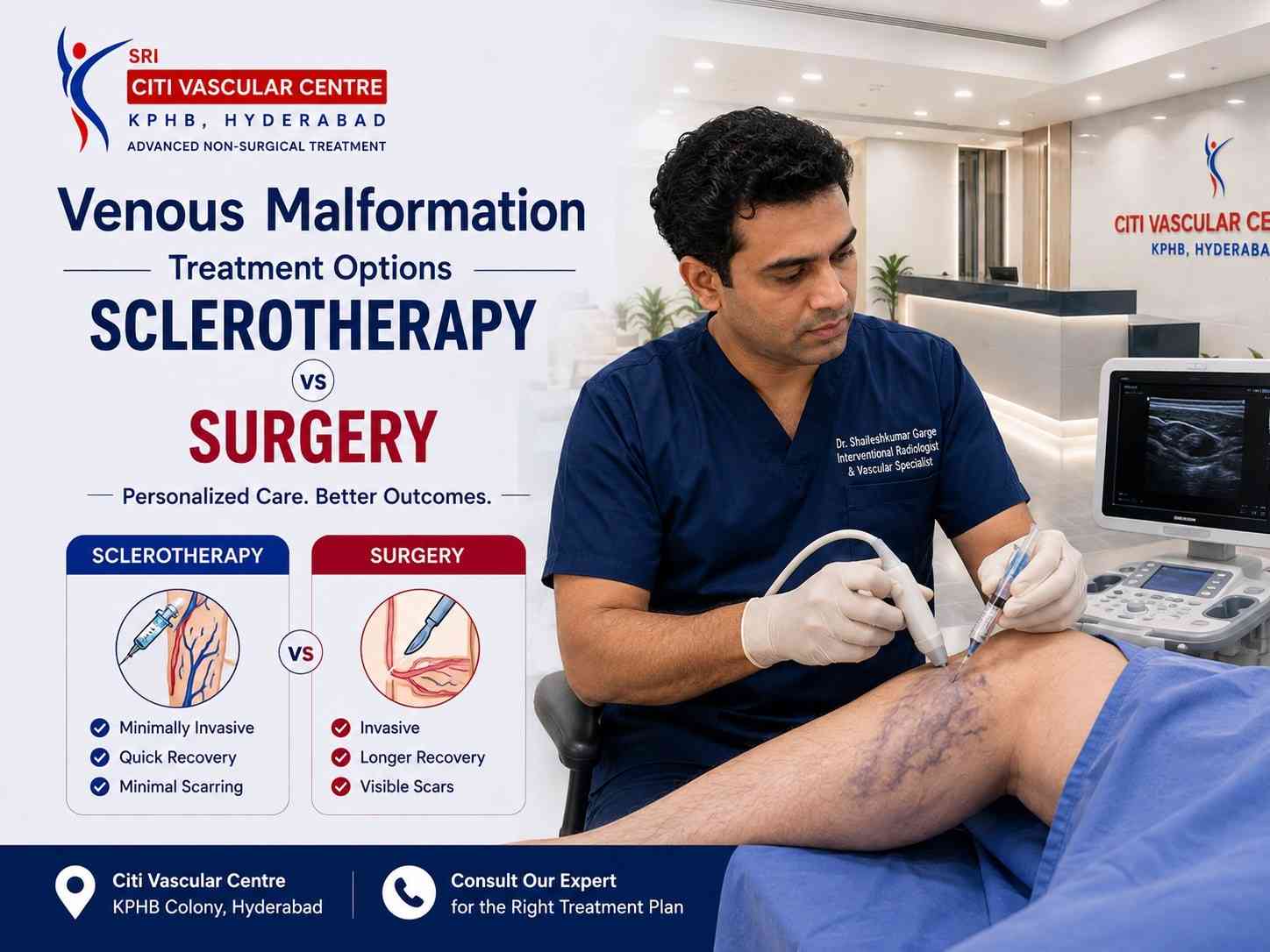
Neither is universally better. For most symptomatic venous malformations, image-guided sclerotherapy is the preferred first-line treatment — minimally invasive, lower blood loss risk, and highly effective for a wide range of VM sizes. Surgery is the right choice for small accessible VMs where complete excision in one operation is achievable, or when sclerotherapy alone cannot adequately address the residual disease. Many complex VMs benefit most from combination treatment: sclerotherapy first, surgery after. Dr. Garge FRCR (UK) | Citi Vascular Centre, KPHB, Hyderabad. Call +91-73375 83901.
When a patient with a symptomatic venous malformation is told that treatment is indicated, two main options typically come onto the table: image-guided sclerotherapy and surgical excision. For many patients — particularly those who have been advised surgery by a non-specialist — the first question is whether sclerotherapy is available, how it compares with surgery, and which is the right choice for their specific VM. This page answers that question with the clinical detail that patients and referring clinicians need.
The short answer is that this is not a simple either-or comparison — it is a clinical decision that depends on the VM's size, location, depth, tissue planes involved, the patient's age and comorbidities, and what outcome the patient is prioritising. For the majority of symptomatic venous malformations, sclerotherapy is the recommended first-line intervention — endorsed by the Society of Interventional Radiology (SIR), CIRSE, and ISSVA guidelines. Surgery is indicated in specific, well-defined clinical situations where sclerotherapy alone is inadequate or inappropriate. And many complex VMs are best managed with a planned combination of both treatments — sclerotherapy to reduce volume and fibrose the channels, followed by surgical excision of the residual and now more surgically manageable fibrous tissue.
This guide provides a complete, balanced, evidence-informed comparison across every dimension that matters: which VMs respond best to each treatment, recovery, cosmetic outcomes, risks, recurrence rates, and the specific clinical situations where combination therapy is the correct approach. For detailed information about the sclerotherapy procedure and surgical technique, see our dedicated pages. For cost comparison, see our VM Cost in Hyderabad page.
Discuss Sclerotherapy vs Surgery at Citi Vascular Centre, KPHB, HyderabadBring your MRI and Doppler USG | Call +91-73375 83901 | WhatsApp 73375 83901 | Mon–Sat 9AM–6PM
Book your Appointment here: | Appoinment: |Call +91-7032508468| Whatsapp:+91-7337583901
|
Feature |
Image-Guided Sclerotherapy |
Surgical Excision |
|
Approach |
Injection of sclerosant into VM channels under USG ± fluoroscopy |
Surgical dissection and removal of VM tissue under general anaesthesia |
|
Incision |
No surgical incision — needle puncture only |
Open surgical incision of variable size depending on VM |
|
Anaesthesia |
Local anaesthesia + IV sedation for most | GA for complex cases |
General anaesthesia — always required |
|
Blood Loss Risk |
Minimal — no VM tissue cut |
Significant — VM channels bleed freely when cut | Transfusion risk in large VMs |
|
Hospital Stay |
Day-care for most sessions |
2–5 nights for major VM excision |
|
Sessions Required |
Multiple (2–5 typically) at 4–8 week intervals |
Usually 1 surgical operation — but completeness limited by blood loss for large VMs |
|
Completeness of Treatment |
Incomplete for very large or diffuse VMs — reduces but rarely eliminates |
Complete for small accessible VMs | Incomplete for large or diffuse VMs |
|
Cosmetic Outcome |
No surgical scar | Gradual external improvement over months |
Surgical scar of variable size | Immediate tissue volume reduction |
|
Recovery |
Days to light activities | 1–2 weeks per session |
2–4 weeks to normal activities | 4–6 weeks to full activity |
|
Nerve Injury Risk |
Low with experienced operator and careful dosing near known nerve locations |
Defined surgical nerve injury risk — varies with VM proximity to nerves |
|
Recurrence |
Possible — partial recurrence in large VMs. Repeat sclerotherapy feasible. |
Possible after incomplete excision. Repeat surgery more complex than repeat sclerotherapy. |
For the complete detailed procedure descriptions, see our dedicated Venous Malformation Sclerotherapy Procedure page and our Venous Malformation Treatment Overview. The brief overview here provides the essential context for comparing the two approaches.
Image-Guided Sclerotherapy
A specialised chemical agent (sclerosant — STS foam, bleomycin, absolute ethanol, or polidocanol) is injected directly into the VM channels under continuous real-time ultrasound guidance, with fluoroscopic confirmation when required. The sclerosant damages the venous lining, causing thrombosis and progressive fibrosis — the VM shrinks over weeks to months. Performed under local anaesthesia + IV sedation for most adult patients. Multiple sessions are usually needed.
Surgical Excision
The VM tissue is surgically dissected and removed under general anaesthesia through a direct incision. For small well-defined VMs, complete excision in a single operation is often achievable. For large or diffuse VMs, complete excision is technically hazardous due to blood loss from the venous malformation channels — which bleed freely when cut. Often combined with pre-operative sclerotherapy to reduce channel blood flow before surgical dissection.
Sclerotherapy is endorsed as the primary interventional treatment for most symptomatic low-flow venous malformations — but it does not work equally well for all VM types. Understanding which VM characteristics predict a better response to sclerotherapy helps set realistic expectations and assists in treatment planning.
|
VM Characteristic |
Sclerotherapy Response |
Why |
|
Small to moderate size (< 6cm) |
Excellent response |
Complete sclerosant coverage of the VM volume achievable within safe dosing limits per session. Higher technical success rate. |
|
Well-defined, localised VM |
Excellent response |
Clear margins allow complete coverage. Less risk of sclerosant tracking into normal tissue. Measurable endpoint on post-procedure imaging. |
|
Intramuscular VM (limb) |
Good response |
Well-contained within muscle planes — sclerosant distributes well within the channel network. Standard USG + fluoroscopy guidance accurate. |
|
Mucosal and submucosal VM (lips, tongue, cheek) |
Good to excellent |
Relatively superficial and accessible. Often responds well to foam sclerotherapy or bleomycin. Avoids disfiguring mucosal surgical scars. |
|
Cystic or predominantly venous lake pattern |
Very good — fewer sessions |
Large venous lakes (expanded venous spaces) fill completely with sclerosant from a single well-placed needle. Higher per-session volume reduction. |
|
VM where surgery carries prohibitive blood loss risk |
Preferred alternative to surgery |
Large, deep, or multi-compartment VMs where surgical dissection would result in unmanageable blood loss — sclerotherapy is the only realistic minimally invasive option. |
|
Large or diffuse VM (> 8–10cm, multi-plane) |
Partial response — may need many sessions |
Cannot always achieve complete elimination in any achievable number of sessions. Realistic goal: significant volume reduction and symptom control, not elimination. |
Surgery is not the preferred first-line approach for most venous malformations — but it is the clinically correct and most effective option in a well-defined set of clinical situations. Understanding when surgery is genuinely the better option — rather than surgery being chosen by default because sclerotherapy is not available — is one of the most important aspects of the clinical decision.
|
Clinical Situation Where Surgery Is Preferred |
Clinical Reasoning |
|
Small, well-defined, accessible VM — complete excision achievable in one operation |
When a VM is small enough (typically < 3–4cm), superficial, and in a location where complete surgical excision is achievable without significant blood loss — surgery can offer definitive single-procedure treatment. No repeated sessions. No waiting months for shrinkage. |
|
Residual or recurrent VM after multiple sclerotherapy sessions |
When adequate sclerotherapy has been performed and significant residual fibrous VM tissue remains — surgical excision of the now-fibrosed and less vascular residual VM is often feasible, safer than operating on the untreated VM, and may achieve complete eradication. |
|
VM containing a defined tissue mass or lipomatous component |
Some VMs have a solid fibrous or lipomatous core that does not respond to sclerotherapy (which treats the venous channels) — surgical removal of this component may be required. |
|
VM causing airway compression requiring immediate volume reduction |
Acute airway compromise from a rapidly expanding VM in the tongue, floor of mouth, or neck requires urgent surgical decompression rather than the gradual volume reduction achievable by sclerotherapy. |
|
Patient preference for definitive single-procedure removal |
Some patients prefer the finality of surgical excision over a series of sclerotherapy sessions extended over many months — a valid personal preference that should be respected when surgical risk is acceptable. |
|
Diagnostic uncertainty — histological examination required |
When VM cannot be confidently distinguished from another soft tissue lesion on imaging alone — surgical excision providing a complete specimen for histopathological examination may be indicated. |
|
VM unsuitable for safe sclerotherapy due to anatomy |
Some VM locations are genuinely technically difficult to access safely with a needle — or the VM's drainage pattern creates unacceptable risk of sclerosant reflux into normal vessels. Surgery may be the safer option. |
The most clinically effective management strategy for many significant venous malformations is not sclerotherapy alone or surgery alone — it is a planned combination of both, used in sequence. This combination approach is endorsed in published international guidelines and is the standard of care for complex VMs at high-volume vascular anomaly centres worldwide. Understanding how the combination works — and why the sequence matters — helps patients engage with their treatment plan.The clinical logic is straightforward. Operating directly on an untreated large venous malformation is one of the most hazardous elective surgical procedures in soft tissue surgery — the VM channels bleed freely when cut, blood loss can be massive and difficult to control, and complete excision is rarely achievable. The same VM that is surgically dangerous before treatment often becomes surgically manageable after sclerotherapy — because the treated channels have thrombosed and fibrosed, the VM is smaller, less vascular, and easier to dissect cleanly from adjacent structures.
MRI and Doppler USG confirm low-flow VM — extent, location, and relationship to adjacent nerves and vessels mapped before any treatment.
Sclerotherapy sessions (typically 2–4) reduce the VM volume by 50–70%, fibrose the venous channels, and convert the highly vascular bleeding VM into a fibrous mass with substantially reduced blood content. D-dimer improves as LIC resolves.
MRI is repeated after the sclerotherapy course — confirming volume reduction, assessing residual VM extent, and planning the surgical approach based on the now-smaller, better-defined residual fibrous mass.
Surgical excision of the residual fibrosed VM — now substantially smaller, less vascular, and better defined than before treatment. Intraoperative blood loss is a fraction of what it would have been operating on the untreated VM. Complete or near-complete excision is much more likely.
Post-operative MRI confirms the extent of excision. Long-term surveillance imaging monitors for any late recurrence in the surgical bed or adjacent tissue.
Who benefits most from combination treatment? Patients with: large (> 6cm) localised VMs that are too large for sclerotherapy alone to achieve adequate reduction | VMs in locations where complete surgical excision without sclerotherapy preparation would risk unacceptable blood loss | Complex head, neck, or craniofacial VMs | Intramuscular VMs in major functional muscle groups. Dr. Garge and the multidisciplinary team discuss the combination approach at consultation for patients where this is the most appropriate plan. Call +91-73375 83901.
Book your Appointment here: | Appoinment: |Call +91-7032508468| Whatsapp:+91-7337583901
|
Recovery Aspect |
After Sclerotherapy Session |
After Surgical Excision |
|
Procedure Day |
1–4 hours observation. Compression dressing. Home same day. Mild-to-moderate pain beginning as local anaesthetic wears off. |
Hospital ward admission. IV drip. Wound drain (often for VM surgery). Recovery from general anaesthesia. |
|
Pain Level — First 48 Hours |
Moderate — inflammatory response at VM site. Paracetamol and prescribed NSAID adequate for most patients. |
Surgical wound pain — stronger analgesics required initially. Drain discomfort. Position-dependent. |
|
Post-Procedure Swelling |
Expected and significant — VM may appear LARGER in Days 3–5 before reducing. This is the therapeutic response, not a complication. |
Surgical oedema around excision site — gradually resolves over 2–3 weeks. |
|
Return to Desk Work |
Day 5–10 for most patients after each session |
2–3 weeks — when wound is comfortable and energy levels allow |
|
Return to Full Activity |
2–3 weeks after each session |
4–6 weeks after major VM excision — wound healing and stamina recovery |
|
Wound Care |
Compression dressing for 24–48 hours. No wound to dress. Tiny needle marks heal within days. |
Wound dressing changes. Suture or staple removal (typically Day 10–14). Drain management. |
|
Number of Recovery Periods |
One recovery period per session × 2–5 sessions — recovery distributed over months |
Single recovery period — but longer and more significant than any single sclerotherapy session |
|
Visible Benefit Timeline |
Gradual improvement over months after each session — maximum at 3–6 months post-course |
Immediate removal of VM volume — though surgical swelling initially obscures the cosmetic improvement |
Cosmetic outcome is one of the most important considerations for patients with visible venous malformations — particularly facial, lip, and cheek VMs in younger patients for whom the impact on appearance has significant psychosocial implications. The comparison between sclerotherapy and surgery on cosmetic outcomes is nuanced.
|
Cosmetic Aspect |
Sclerotherapy |
Surgical Excision |
|
Entry Point Marks |
Tiny needle marks (1–2mm) — heal to invisible within days. |
Surgical incision — variable in length and prominence depending on VM size and location. Usually planned in skin crease where possible. |
|
Visible Scar |
No surgical scar in most cases |
Surgical scar of variable size — fades over 12–24 months but remains visible in most patients to some degree |
|
Speed of Cosmetic Improvement |
Gradual — 50–70% VM volume reduction over 3–6 months per session. Multi-session improvement cumulative. |
Immediate reduction in VM volume visible at surgery — though post-surgical swelling initially delays cosmetic result. |
|
Completeness of External Appearance Change |
High satisfaction for moderate VMs. Very large or diffuse VMs may achieve significant but not complete cosmetic improvement even after multiple sessions. |
High for small accessible VMs — complete excision means complete volume removal. Less complete for large or deep VMs where excision is partial. |
|
Risk of Cosmetic Complication |
Skin blistering or superficial ulceration — more common with absolute ethanol in very superficial VMs. Managed with wound dressing. |
Wound dehiscence, hypertrophic scar, keloid (particularly in high-risk anatomical locations and patients with keloid tendency), visible scar distortion. |
|
Best for Cosmetically Sensitive Locations |
Generally preferred for facial, lip, and oral mucosal VMs — avoids surgical scar in highly visible areas |
May be preferred for VMs in areas where skin crease incisions are well-concealed — e.g. behind ear, in skin folds |
Cosmetic outcome and surgical approach planning: When surgery is performed for VM, the incision placement is planned carefully to minimise visible scarring — using natural skin creases and hidden locations where possible. At Citi Vascular Centre, for patients who require surgical excision, Dr. Garge coordinates with plastic surgeons or maxillofacial surgeons experienced in VM surgery to optimise both the oncological completeness and the cosmetic outcome of the incision.
|
Risk |
Sclerotherapy Risk Profile |
Surgical Excision Risk Profile |
|
Major blood loss |
Very low — sclerosant does not cut tissue. No significant blood loss from the procedure. |
Significant risk — VM channels bleed freely when dissected. Risk of major haemorrhage and transfusion requirement for large VMs. Most dangerous unique risk of VM surgery. |
|
Post-procedure pain and swelling |
Common and expected — 1–2 weeks per session. Managed with analgesics. |
Surgical wound pain — more significant than sclerotherapy per session. Managed with stronger analgesics initially. |
|
Nerve injury |
Risk with VMs adjacent to named nerves. Minimised by hydrodissection and careful dosing. Experienced operator essential. |
Surgical nerve injury risk — variable depending on VM proximity to named nerves. More difficult to 'pause' intraoperatively than during injection. |
|
Skin damage / wound complication |
Skin blistering or superficial ulceration — uncommon. More frequent with absolute ethanol in very superficial VMs. |
Wound dehiscence, infection, delayed healing — particularly if post-operative haematoma develops. |
|
Anaesthetic risks |
Local anaesthesia risks minimal. IV sedation: very low risk. GA (when required): moderate risk depending on patient comorbidities. |
General anaesthesia required — always carries intubation risk, PONV, and cardiovascular risk — quantified specifically by pre-anaesthetic assessment. Greater risk in elderly patients or those with comorbidities. |
|
Deep vein thrombosis |
Rare — primarily with large VMs and absolute ethanol sclerotherapy. Anticoagulation monitoring reduces risk. |
Post-operative DVT risk — particularly for lower limb VM surgery where immobility is greater. Routine anticoagulation prophylaxis. |
|
Infection |
Rare — < 1%. Sterile technique prevents. |
Surgical site infection risk — 1–3% for clean elective surgery. Antibiotic prophylaxis standard. |
|
Incomplete treatment / residual VM |
Expected for large VMs — each session reduces, rarely eliminates. Multiple sessions planned. |
More consequential — if surgery leaves significant residual VM, reoperation is technically more difficult than primary surgery. |
Both sclerotherapy and surgery carry a risk of VM recurrence — and understanding what 'recurrence' means in this context, and how the long-term outlook differs between the two approaches, helps patients plan for realistic long-term management.
|
Long-Term Consideration |
Sclerotherapy vs Surgery — What the Evidence Shows |
|
Can the VM come back after treatment? |
Yes — for both approaches. The underlying developmental anomaly of venous tissue remains. New dilated channels can develop adjacent to treated tissue over time. Recurrence is more clinically significant than residual disease — but both are managed similarly. |
|
Recurrence after sclerotherapy |
Partial recurrence is possible — particularly in large or diffuse VMs where the sclerotherapy course produced significant but not complete fibrosing. Repeat sclerotherapy is safe, effective, and follows the same technical approach as original sessions. |
|
Recurrence after surgery |
Recurrence after incomplete surgical excision is reported — particularly for large or diffuse VMs where completeness of excision was limited by blood loss or proximity to important structures. Reoperation is technically more demanding than primary surgery due to post-surgical scarring. |
|
Which has better long-term durability — sclerotherapy or surgery? |
For small accessible VMs where complete surgical excision is achievable — surgery provides the most durable outcome. For larger or deeper VMs — neither approach provides a completely reliable long-term cure, and ongoing surveillance with retreatment as needed is the realistic long-term plan for both sclerotherapy and surgery patients. |
|
How is recurrence managed? |
After sclerotherapy: repeat sclerotherapy sessions — straightforward and technically similar to original treatment. After surgery: combination of repeat sclerotherapy (to reduce the recurrent VM volume) and possible repeat surgical excision if the recurrent lesion is technically accessible. |
|
Surveillance imaging after treatment |
Long-term annual or biennial MRI surveillance is recommended after either treatment for significant VMs — to detect recurrence early, at a stage when retreatment is more effective and less complex than waiting for symptomatic recurrence. |
This flowchart reflects the clinical decision-making logic at Citi Vascular Centre, KPHB — not a substitute for Dr. Garge's assessment of your individual case, but a framework to help patients understand how the recommendation is arrived at.
|
▼ |
VM confirmed as low-flow on Doppler USG — no arterial signal. MRI maps full 3D extent. D-dimer and coagulation checked. |
|
? |
Is the VM small (< 3–4cm), well-defined, superficial, and in a location where surgical excision is achievable with low blood loss risk? |
|
YES |
Consider single-operation surgical excision if patient accepts GA risk and surgical scar. Alternatively, sclerotherapy as first-line if patient prefers to avoid surgery. Both are appropriate — patient preference informed by specialist discussion. |
|
NO |
VM is large, deep, diffuse, intramuscular, or in a location where surgical blood loss would be hazardous → Sclerotherapy first-line. Multiple sessions planned. MRI between sessions monitors response. |
|
▼ |
After 2–4 sclerotherapy sessions — MRI reassessment. Significant residual VM remains but is now fibrosed and smaller. |
|
? |
Is residual VM causing ongoing symptoms? Is surgical excision of residual disease technically feasible after fibrosis? |
|
YES |
Consider surgical excision of residual fibrosed VM — now safer, more technically feasible, and more likely to be complete than operating on the original untreated VM. |
|
NO |
Patient satisfied with symptom improvement from sclerotherapy alone → Continue long-term surveillance with periodic imaging. Retreat if recurrence detected. |
|
▼ |
Long-term: annual or biennial MRI surveillance. Retreatment — sclerotherapy or surgery — if significant recurrence detected. |
|
|
Myth |
Fact |
|
1 |
Surgery is the most effective treatment for venous malformation. |
For most VMs, sclerotherapy is the preferred first-line intervention. Surgery carries higher blood loss risk for large VMs and is often incomplete without prior sclerotherapy to reduce VM vascularity. |
|
2 |
Sclerotherapy only works for very small venous malformations. |
Sclerotherapy is effective across a wide range of VM sizes and can achieve meaningful volume reduction in large VMs over multiple sessions, even when complete elimination is not achievable. |
|
3 |
Surgery cures venous malformation permanently — it won't come back. |
Surgical excision reduces the VM volume and may achieve complete removal for small accessible lesions — but recurrence is possible after incomplete excision, and the underlying vascular anomaly tendency means new channels can develop over time. |
|
4 |
Sclerotherapy and surgery are alternative choices — you do one or the other. |
Many complex VMs benefit most from planned combination treatment: sclerotherapy first to reduce volume and fibrose the channels, followed by surgical excision of residual fibrosed tissue — often the safest and most complete approach overall. |
|
5 |
Sclerotherapy leaves no scar but surgery always leaves a very visible scar. |
Sclerotherapy leaves no surgical scar, but may cause skin blistering in very superficial VMs. Surgical scars vary enormously by location, approach, and surgeon skill — carefully placed incisions in skin creases often produce acceptable cosmetic outcomes. |
|
6 |
General anaesthesia is required for sclerotherapy. |
Most adult VM sclerotherapy sessions at Citi Vascular Centre are performed under local anaesthesia and IV sedation — not general anaesthesia. GA is needed for children and complex head/neck VM sessions. |
Q1: Is sclerotherapy better than surgery for venous malformation?
For most symptomatic venous malformations, sclerotherapy is the preferred first-line interventional treatment — endorsed by SIR, CIRSE, and ISSVA guidelines. It avoids the significant blood loss risk of VM surgery, preserves surrounding tissue, leaves no surgical scar, and is effective for a wide range of VM sizes. Surgery is the right choice for small accessible VMs where complete surgical excision in one operation is achievable, or as a planned second step after sclerotherapy volume reduction in complex cases.
Q2: Can venous malformation be treated without surgery?
Yes — for the majority of venous malformations, surgery is not required. Image-guided sclerotherapy effectively reduces VM volume, relieves pain, and improves function in most patients through 2–5 treatment sessions. Small VMs may respond to a single session. Very large or diffuse VMs may benefit from combination sclerotherapy + surgery, but even in these cases, sclerotherapy is the first step. Surgery alone — without prior sclerotherapy — is appropriate only for small, surgically accessible VMs.
Q3: Which venous malformations are too large for sclerotherapy?
There is no absolute size limit that makes sclerotherapy impossible — but very large (> 8–10cm) or diffuse multi-compartment VMs present challenges because: the sclerosant volume safe per session is limited; complete coverage of a very large VM in one session is not achievable; and many sessions may only achieve partial reduction. For these VMs, staged sclerotherapy reduces the VM to a more manageable size, after which surgical excision of the residual fibrosed tissue is often recommended as a combined approach.
Q4: What happens if sclerotherapy does not work well enough?
If sclerotherapy achieves adequate symptom control and volume reduction — treatment continues with periodic surveillance. If significant residual VM remains after a full sclerotherapy course, several options exist: additional sclerotherapy sessions; surgical excision of the residual fibrosed VM (which is now safer and more technically feasible than operating on the untreated VM); or observation if the residual volume is asymptomatic. Dr. Garge reviews the MRI response at each follow-up and discusses the management plan at every step.
Q5: What is combination treatment for venous malformation?
Combination treatment uses sclerotherapy first, followed by surgical excision of residual VM. The approach is used for larger or complex VMs: 2–4 sclerotherapy sessions reduce the VM volume by 50–70% and fibrose the channels — making the VM smaller, less vascular, and safer to excise. Post-sclerotherapy surgical excision of the residual fibrosed mass is technically safer and more complete than operating on the untreated VM. This is often the best long-term outcome strategy for significant VMs.
Q6: Does sclerotherapy leave a scar?
No surgical scar — image-guided sclerotherapy requires only needle punctures (1–2mm) that heal to invisible marks within days. There is no incision, no wound, and no sutures. This is one of the most significant cosmetic advantages of sclerotherapy over surgical excision — particularly for facial, lip, or mucosal VMs where surgical scars would be visible and potentially disfiguring. Skin blistering from aggressive sclerosants is an uncommon complication, not a routine outcome.
Q7: Can a venous malformation come back after surgery?
Yes — recurrence after VM surgery is possible, particularly when complete excision is not achieved (which is common for large or deep VMs). Post-surgical scarring makes reoperation technically more challenging than primary surgery. Recurrent VMs after surgery can usually be managed with sclerotherapy in the first instance, followed by repeat surgical excision if the recurrent lesion is accessible. Long-term MRI surveillance after either surgery or sclerotherapy is recommended to detect recurrence early.
Q8: Which treatment has a faster recovery — sclerotherapy or surgery?
Individual session recovery from sclerotherapy (1–2 weeks per session) is faster than surgical excision recovery (2–4 weeks). However, sclerotherapy requires multiple recovery periods over months for a complete multi-session course, whereas surgery involves one longer recovery. Most patients prefer the shorter individual recovery of sclerotherapy — particularly because they can return to work and normal activities between sessions. The choice depends partly on how the total disruption of multiple short recoveries compares with one longer recovery for the patient's specific situation.
Q9: Is general anaesthesia always required for venous malformation surgery?
Yes — surgical excision of a venous malformation always requires general anaesthesia. Surgery requires the patient to be completely still, the airway to be secured, and blood pressure management to minimise surgical blood loss. There are no safe options for VM surgery under local anaesthesia alone. This is in contrast to sclerotherapy, where most adult sessions are performed under local anaesthesia and IV sedation — with general anaesthesia reserved for children and complex cases.
Q10: Which treatment is better for facial venous malformations?
For most facial venous malformations, sclerotherapy is preferred over primary surgery for several reasons: it avoids a surgical scar in a highly visible location, can treat deep components that cannot be safely excised without facial nerve risk, and produces gradual cosmetic improvement over multiple sessions. Surgical excision of facial VMs carries significant risks to facial nerve branches and produces visible scars. Combined sclerotherapy + limited surgery is sometimes used for persistent residual facial VM after adequate sclerotherapy response.
Q11: How do I know which treatment is right for my venous malformation?
The right treatment for your VM is determined by your specific clinical picture — VM size, location, depth, MRI extent, symptoms, your age, your surgical fitness, and your personal priorities around scarring, recovery time, and definitive removal. No online comparison can substitute for a specialist review of your imaging and clinical assessment. Bring your MRI and Doppler USG report to a consultation with Dr. Garge at Citi Vascular Centre, KPHB, for a specific, imaging-based recommendation. Call +91-73375 83901.
Q12: Who performs venous malformation sclerotherapy in Hyderabad?
Image-guided sclerotherapy for venous malformation should be performed by an Interventional Radiologist with specific training in vascular anomaly management. At Citi Vascular Centre, KPHB Colony, Hyderabad, all VM sclerotherapy is performed by Dr. Shaileshkumar Garge — FRCR (UK), FNVIR (CMC Vellore), EBIR (Spain) — using ISSVA-aligned classification, combined USG + fluoroscopic guidance, and personalised sclerosant selection. Surgical cases are coordinated with experienced vascular and maxillofacial surgeons. Call +91-73375 83901.
EVIDENCE-BASED REFERENCES
|
Source |
Reference |
|
ISSVA Classification |
International Society for the Study of Vascular Anomalies (ISSVA). Classification of Vascular Anomalies. 2018. issva.org. — Basis for VM classification as low-flow malformations and sclerotherapy as primary treatment modality. |
|
SIR Clinical Guidelines |
Society of Interventional Radiology (SIR). Clinical Practice Guidelines — Image-Guided Management of Venous Malformations. sirweb.org. — Recommends sclerotherapy as first-line for most symptomatic low-flow VMs. |
|
CIRSE Standards |
Cardiovascular and Interventional Radiological Society of Europe (CIRSE). Standards of Practice: Sclerotherapy for Venous Malformations. Cardiovasc Intervent Radiol. — Defines sclerotherapy indications, contraindications, and combination approach with surgery. |
|
Combination Treatment |
Dompmartin A et al. Management of venous malformations: the sclerotherapy-first and combined approach. Ann Vasc Surg. — Evidence base for staged sclerotherapy + surgery as optimal strategy for complex VMs. |
|
Recurrence After Treatment |
Lee BB. Consensus on recurrence of venous malformation after treatment: sclerotherapy versus surgical excision — systematic review. J Vasc Surg. — Long-term recurrence rates and management strategies after each treatment modality. |
LOCATION — VM SCLEROTHERAPY vs SURGERY IN HYDERABAD
Citi Vascular Centre, KPHB Colony, Road No. 1, Hyderabad — VM treatment assessment for patients from:
Kukatpally and KPHB — 5 min
Miyapur and Bachupally — 10 min
Hitech City, Madhapur and Ameerpet — 20 min
Gachibowli and Banjara Hills — 25 min
Secunderabad and Begumpet — 25 min
Kompally, Medchal and Alwal — 20–25 min
Telangana and Andhra Pradesh — outstation patients welcome
|
Centre |
Contact |
Hours |
|
Citi Vascular Centre |
+91-73375 83901 |
KPHB Colony, Road No. 1, Hyderabad, Telangana 500072 | Mon–Sat 9AM–6PM |
|
|
73375 83901 |
Bring MRI and Doppler USG | Same-week appointments | Outstation patients welcome |
KEY TAKEAWAYS
Neither sclerotherapy nor surgery is universally better — the right choice depends on VM size, location, depth, patient fitness, and personal priorities
Sclerotherapy is preferred first-line for most symptomatic VMs — SIR, CIRSE, ISSVA guidelines endorsed. Lower blood loss risk, no surgical scar, wide range of appropriate VM sizes
Surgery is preferred for: small accessible VMs achievable in one operation | residual VM after sclerotherapy | acute airway compromise | diagnostic uncertainty requiring histology
Combination treatment (sclerotherapy then surgery) is the optimal strategy for many large or complex VMs — sclerotherapy reduces volume and fibroses channels, making subsequent surgery safer and more complete
Post-sclerotherapy swelling (VM appears larger in Days 3–5) is the therapeutic response — not failure. Maximum response assessed by MRI at 2–3 months per session.
Dr. Shaileshkumar Garge FRCR (UK) | Citi Vascular Centre, KPHB | +91-73375 83901 | WhatsApp 73375 83901 | Bring MRI + Doppler USG | Mon–Sat 9AM–6PM
SUMMARY
Venous malformation sclerotherapy and surgical excision are complementary — not competing — treatments. The clinical decision between them, or the decision to combine them, depends entirely on the individual patient's VM: its size, its location, how deep it lies, its relationship to nerves and vessels, how symptomatic it is, and what the patient's priorities are. For most patients with symptomatic venous malformations, image-guided sclerotherapy is the evidence-based preferred first-line interventional option — reducing VM volume over multiple sessions without surgical blood loss risk, without surgical scar, and with recovery measured in days per session rather than weeks. Surgery adds the most value for small, surgically accessible VMs where one operation can achieve complete excision, or as a planned second step after sclerotherapy has made a large VM safer and more completable surgically.
The most important message of this comparison page is that the answer to 'sclerotherapy vs surgery' for venous malformation is almost always 'it depends on your specific VM' — and that answer can only be given after a specialist reviews your Doppler ultrasound and MRI. At Citi Vascular Centre, KPHB Colony, Hyderabad, Dr. Shaileshkumar Garge provides sclerotherapy, coordinates surgical referral, and plans combination approaches — giving every patient the full range of options and a recommendation based on their imaging, not on which treatment the centre happens to offer. Bring your MRI and Doppler USG to a consultation. Call +91-73375 83901 or WhatsApp 73375 83901.
Sclerotherapy or Surgery? Get an Honest Recommendation for Your Venous Malformation
Bring: MRI Report + Disc | Doppler USG | D-dimer | Any previous treatment records
Dr. Shaileshkumar Garge | FRCR (UK) | FNVIR (CMC Vellore) | EBIR (Spain) | 12+ Years | 15,000+ Procedures
Call +91-73375 83901 | WhatsApp 73375 83901 | citivascularcentre.com
Citi Vascular Centre, KPHB Colony, Hyderabad | Mon–Sat 9AM–6PM | Outstation Welcome
Book your Appointment here: | Appoinment: |Call +91-7032508468| Whatsapp:+91-7337583901