LAST MEDICALLY REVIEWED:
August 2026 — Dr. Shaileshkumar Garge
Citi Vascular Hospital, KPHB Colony, Road No. 1, Hyderabad, Telangana 500072
|
QUICK ANSWER What Is a Venous Malformation and How Is It Treated in Hyderabad? A venous malformation is a congenital (present-from-birth) abnormality of veins — soft, compressible, blue-purple in colour, and present in any part of the body. It is not cancer, not an infection, and does not go away on its own. Treatment — usually image-guided sclerotherapy, sometimes combined with surgery — reduces size, relieves pain, and improves function. Dr. Garge FRCR (UK) | Citi Vascular Centre, KPHB, Hyderabad. Call +91-73375 83901. |
A venous malformation is one of the more common types of vascular malformation — and one of the most frequently misunderstood. Patients are often told for years that the soft, bluish lump somewhere on their body is a 'haemangioma', a 'blood blister', or a harmless fatty lump, before an imaging study reveals what it actually is: a cluster of abnormally dilated, poorly formed venous channels that have been there since birth, filled with slow-moving venous blood, and gradually enlarging over time.
Venous malformations are not tumours, not infections, not varicose veins, and not lymphangiomas — though they can occasionally be confused with all of these on clinical examination alone. They are developmental vascular anomalies — errors in the formation of the venous system during fetal development — and they belong to the category of low-flow vascular malformations in the internationally recognised ISSVA classification. Unlike haemangiomas (which typically involute in childhood), venous malformations do not go away on their own and tend to gradually increase in size over the years.
This guide explains what venous malformations are, what causes them, the different types, how they present clinically, how they are accurately diagnosed using MRI and Doppler ultrasound, and what the treatment options include — from observation through image-guided sclerotherapy to surgery. For detailed information about the sclerotherapy procedure itself, see our dedicated Sclerotherapy for Venous Malformation page.
Venous Malformation Consultation — Citi Vascular Centre, KPHB, Hyderabad
Call +91-73375 83901 | WhatsApp 73375 83901 | citivascularcentre.com | Mon–Sat 9AM–6PM
|
Feature |
Detail |
|
What It Is |
Congenital slow-flow vascular malformation — abnormally dilated venous channels present since birth |
|
Classification |
Low-flow vascular malformation (ISSVA 2018) — distinct from haemangioma, arteriovenous malformation, and lymphatic malformation |
|
Is It Cancer? |
No — venous malformation is a benign developmental vascular anomaly. Not a tumour. |
|
Does It Go Away? |
No — venous malformations do not spontaneously resolve. They tend to slowly enlarge over years. |
|
Common Locations |
Face, neck, tongue, lip, limbs, trunk — virtually anywhere in the body or within organs |
|
How It Looks/Feels |
Soft, compressible, blue-purple discolouration — enlarges when the affected part is in a dependent position or with Valsalva manoeuvre |
|
Diagnosis |
Doppler ultrasound (first-line) + MRI (definitive extent mapping) | D-dimer (elevated in large VMs) |
|
Treatment Options |
Observation | Compression garments | Image-guided sclerotherapy | Surgery | Laser (superficial cases) |
|
First-Line Treatment |
Image-guided sclerotherapy — most symptomatic VMs are treated with sclerotherapy as first-line intervention |
|
Who Treats It? |
Interventional Radiologist — for sclerotherapy | Vascular Surgeon — for surgical excision | Combined MDT for complex cases |
A venous malformation (VM) is a developmental vascular anomaly — an error that occurs during the formation of the venous system in the developing fetus. Instead of forming normal, well-defined veins with proper smooth muscle walls, a cluster of abnormally dilated, thin-walled, poorly contractile venous channels forms within the tissue. These channels are filled with venous blood that moves very slowly — creating the characteristic low-flow vascular malformation that can be confirmed by Doppler ultrasound.
Venous malformations are present from birth — they are congenital lesions, though they may not be immediately visible or symptomatic at birth. Many venous malformations are first noticed during childhood, adolescence, or early adulthood when they become more apparent — often due to hormonal changes (puberty, pregnancy) that cause enlargement, or when a minor trauma or thrombotic episode draws attention to a previously unnoticed lump. Some remain entirely undiagnosed until an unrelated imaging study is performed.
The key distinction that is most important for patients to understand is the difference between a venous malformation and a haemangioma — a distinction that has significant clinical implications. Haemangiomas (more accurately infantile haemangiomas) are vascular tumours that typically appear in infancy, grow rapidly in the first year of life, and then involute (shrink) spontaneously over subsequent years. Venous malformations, by contrast, do not spontaneously resolve, grow slowly and progressively over years, and require active management when symptomatic. The ISSVA (International Society for the Study of Vascular Anomalies) 2018 classification formally separates vascular tumours (including haemangiomas) from vascular malformations — and it is the malformation group that includes venous malformations.
Important: The term 'haemangioma' is commonly — and incorrectly — applied to venous malformations by patients and even some clinicians. If you have been told your lump is a 'haemangioma', ask specifically whether imaging has been done and whether the diagnosis has been confirmed by a vascular specialist. Many so-called 'haemangiomas' in adults are actually venous malformations requiring different management. Call +91-73375 83901.
Venous malformations are caused by errors in vascular morphogenesis — the complex cellular process by which blood vessels form and differentiate during fetal development. The exact cause at the molecular level has been increasingly characterised by genetic research over the past two decades.
|
Cause / Factor |
What the Evidence Shows |
|
Sporadic somatic mutation |
Most venous malformations occur as sporadic (non-inherited) mutations that happen during fetal vascular development — not passed from parent to child. The TIE2/TEK receptor tyrosine kinase gene mutation is the most commonly identified somatic driver mutation in sporadic VMs. |
|
Inherited / familial forms |
A minority of VMs are inherited. Mutations in the TEK gene (autosomal dominant) cause familial cutaneous and mucosal venous malformations (VMCM) — multiple small cutaneous VMs appearing in several family members. Hereditary haemorrhagic telangiectasia (HHT) is a related condition. |
|
Vascular channel wall deficiency |
VM channels have abnormal smooth muscle cells and thin walls — unable to contract or maintain normal venous tone. This leads to progressive dilation and pooling of venous blood within the abnormal channels. |
|
Not caused by trauma or infection |
Venous malformations are not caused by injury, infection, medication, or lifestyle factors. They are structural developmental anomalies — no patient behaviour causes or prevents them. |
|
Hormonal influence |
While not a cause, hormonal changes at puberty and pregnancy can accelerate VM enlargement. Oestrogen and progesterone appear to promote growth of venous malformation tissue — which is why many VMs become more symptomatic or more apparent during these hormonal transitions. |
|
Not preventable |
Because VMs arise from fetal developmental errors — most of which are random somatic mutations — they cannot currently be prevented. Early accurate diagnosis is more clinically valuable than any preventive measure. |
Venous malformations vary widely in their clinical behaviour, severity, and management requirements depending on their size, extent, depth, location, and whether they are isolated or part of a syndromic condition. Understanding the type of VM a patient has is essential for treatment planning.
|
Type |
Description |
|
Localised venous malformation |
A single, well-defined cluster of abnormal venous channels — most commonly in the subcutaneous tissue, muscle, or mucosa. The most common type seen in clinical practice. Often amenable to sclerotherapy in one or more sessions. |
|
Diffuse / infiltrating venous malformation |
Spreads through multiple tissue planes — skin, subcutaneous fat, muscle, sometimes bone. Less amenable to complete treatment. Management focuses on symptom control and reducing the most symptomatic components. |
|
Cutaneous / mucocutaneous VM |
Involves the skin surface and mucous membranes — lips, tongue, cheek, gums. Blue-purple discolouration visible on the skin or mucosa. May respond well to laser therapy (superficial cutaneous component) and sclerotherapy (deeper components). |
|
Intramuscular VM |
Lies within or between muscle groups — commonly in the limbs. Often presents with exercise-induced pain and swelling. Risk of thrombosis and phleboliths (calcified clots within VM channels). Sclerotherapy is the primary treatment. |
|
Intraosseous VM |
Within bone — mandible (jaw) most common. Often an incidental finding on dental or maxillofacial imaging. Can cause pain, swelling, and pathological fracture if untreated. |
|
Visceral / deep organ VM |
Within visceral organs — liver, GI tract (Blue Rubber Bleb Nevus Syndrome), spleen. May cause internal bleeding, obstruction, or organ dysfunction depending on location. |
|
Syndromic venous malformation |
Part of a named syndrome: Klippel-Trenaunay Syndrome (VM + lymphatic malformation + capillary malformation + limb overgrowth), Blue Rubber Bleb Nevus Syndrome (BRBN — multiple GI and cutaneous VMs), or VMCM (familial multiple cutaneous VMs). |
The clinical presentation of a venous malformation varies significantly depending on its location, size, and depth. Some venous malformations remain entirely asymptomatic throughout life and are discovered incidentally on imaging. Others cause significant pain, functional limitation, and cosmetic concern that warrants active treatment.
|
Symptom |
How It Presents |
What Causes It |
|
Soft compressible swelling |
A bluish-purple, soft lump that compresses easily under finger pressure and refills when pressure is released |
Slow-flow venous blood within dilated channels — pressure empties the channels, which refill passively from venous blood |
|
Positional enlargement |
The lump visibly enlarges when the affected limb is held in a dependent position (arm hanging down, leg hanging off the bed) or during straining |
Venous blood pools within the malformation channels under gravity or increased venous pressure |
|
Pain and tenderness |
Aching, throbbing, or sharp pain — often worse after prolonged activity, at the end of the day, or after maintaining the dependent position |
Thrombosis within malformation channels (clot formation) is the most common cause of acute pain. Pressure on adjacent structures also contributes. |
|
Acute painful episodes |
Sudden onset of severe localised pain and tenderness — may be accompanied by local warmth and swelling |
Acute intraluminal thrombosis (clotting within the VM channels) — a spontaneous but very painful event common in VMs |
|
Blue-purple skin discolouration |
Bluish discolouration of the overlying skin — may be the only visible sign of a superficial VM |
Venous blood showing through thin overlying skin or mucosal tissue within the superficial VM channels |
|
Functional limitation |
Reduced range of movement in a limb or joint; difficulty swallowing or speaking (oropharyngeal VM); cosmetic disfigurement |
VM within or adjacent to a joint, muscle, or critical structure causing mechanical interference |
|
Palpable phleboliths |
Small, hard, round nodules palpable within the lump |
Phleboliths are calcified old thrombi that have hardened within the VM channels — their presence on X-ray or CT is almost pathognomonic for venous malformation |
|
⚠ |
Rapidly expanding painful swelling in the face or neck |
Rapidly enlarging VMs in the face, floor of mouth, or neck can cause airway compression — a medical emergency requiring urgent specialist assessment. |
|
⚠ |
Difficulty breathing or swallowing |
Oropharyngeal, tongue, or floor-of-mouth VMs that enlarge rapidly can compromise the airway — seek immediate medical attention. |
|
⚠ |
Sudden severe increase in pain |
Acute intravascular thrombosis within a VM causes sudden severe pain. Not life-threatening but requires specialist assessment and anticoagulation management. |
|
⚠ |
Uncontrolled bleeding from a VM |
VMs in the GI tract, airway mucosa, or soft tissue can bleed. Recurrent or uncontrolled bleeding requires urgent interventional or surgical management. |
|
⚠ |
Eye involvement — proptosis, vision change |
Orbital VMs can compress the optic nerve — any visual change or eye prominence associated with a known periorbital VM warrants urgent ophthalmology and IR review. |
Accurate diagnosis of a venous malformation is essential before any treatment can be planned — and it requires a combination of clinical assessment and specific imaging. For many venous malformations, the clinical picture (soft, compressible, blue-purple, positionally variable lump) strongly suggests the diagnosis — but imaging is always required to confirm the diagnosis, characterise the extent, and plan the treatment approach.
|
Diagnostic Step |
What It Provides |
|
Clinical examination |
Assessment of lump size, compressibility, colour, positional change, palpable phleboliths, pain on palpation, relationship to adjacent structures. A highly characteristic clinical picture for venous malformation — but imaging confirmation is always required. |
|
Doppler Ultrasound (First-Line) |
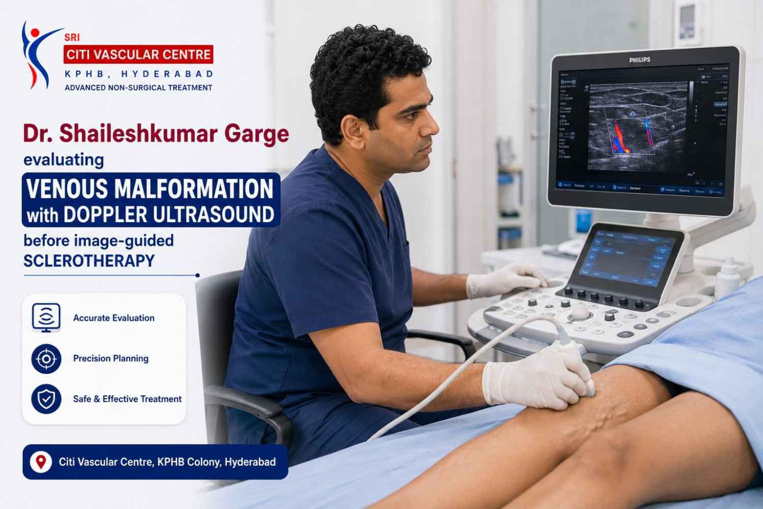
High-resolution Doppler ultrasound is typically the first imaging modality — confirms the vascular nature of the lump, demonstrates the characteristic low-flow venous pattern (no arterial waveform — distinguishing VM from AVM), shows phleboliths as echogenic foci with acoustic shadowing, and assesses compressibility in real time. Performed by Dr. Garge personally at Citi Vascular Centre, KPHB. |
|
MRI — Definitive Extent Mapping |
MRI is the gold standard imaging investigation for venous malformations. T2-weighted MRI demonstrates VMs as high-signal (bright) lesions — because the slow-flowing venous blood produces very high T2 signal. MRI defines the three-dimensional extent of the VM, its relationship to adjacent nerves, vessels, bones, and organs, and guides treatment planning. Gadolinium contrast is used to assess post-treatment response. |
|
D-dimer blood test |
Elevated D-dimer occurs in approximately 40–50% of patients with large venous malformations — due to localised intravascular coagulopathy (LIC) within the slow-flowing VM. Very elevated D-dimer before treatment is a risk factor for more significant coagulopathy during sclerotherapy. Checked as standard before any intervention. |
|
Coagulation profile |
Fibrinogen, PT, APTT, platelets — checked in patients with large VMs before any intervention. Deranged coagulation increases the risk of post-procedure bleeding or coagulopathy flare. |
|
X-ray / CT (selected cases) |
Plain X-ray may demonstrate phleboliths — pathognomonic for VM. CT is useful for VMs with bony involvement (intraosseous VM) and for operative planning. CT angiography is sometimes used to confirm low-flow pattern and exclude arterial component. |
|
Biopsy — rarely needed |
Tissue biopsy is generally NOT required for venous malformation diagnosis and carries a significant bleeding risk from the venous channels. Diagnosis is established by clinical assessment and imaging in most cases. |
|
Why MRI Matters for Venous Malformation T2 MRI maps the full 3D extent of the VM — defining deep components that are not palpable on examination, relationships to nerves and vessels, and the volume to be treated. Without MRI, treatment planning is incomplete. |
Why Doppler Ultrasound Matters Doppler USG distinguishes low-flow VM (no arterial signal) from high-flow arteriovenous malformation (AVM) — a critical distinction as AVM requires completely different treatment. Confirming low-flow status prevents dangerous treatment errors. |
Not every venous malformation requires active treatment. Small, asymptomatic venous malformations in locations where they are not causing functional problems, cosmetic concern, or progressive enlargement can be safely monitored with periodic imaging. The decision to treat is always based on a balanced assessment of the clinical picture — symptoms, size, location, rate of growth, and the patient's individual priorities and expectations.
|
Treatment Is Usually Indicated When |
Observation May Be Appropriate When |
|
Pain that affects quality of life or daily activities |
Small, stable VM with no or minimal symptoms |
|
Acute thrombotic episodes causing recurrent severe pain |
Patient is comfortable with monitoring and understands the natural history |
|
Functional limitation — joint range, swallowing, speech, vision |
VM is in a location where treatment risks outweigh potential benefits |
|
Cosmetic disfigurement causing significant distress |
Very young child where delayed treatment until a more cooperative age is clinically appropriate |
|
VM involving the airway, orbit, or other vital structure |
Asymptomatic incidental finding on imaging with no functional or cosmetic impact |
|
Progressive enlargement on serial imaging |
Risk of complications from treatment exceeds the current symptom burden |
|
GI VM causing bleeding |
Patient chooses observation after being fully informed of all options |
Venous malformation management has evolved significantly over the past two decades. What was once treated almost exclusively by surgery — with its attendant risks of blood loss, incomplete excision, scarring, and recurrence — is now more commonly treated by image-guided sclerotherapy as the primary modality, with surgery reserved for selected cases or used as an adjunct after sclerotherapy has reduced the VM volume. Understanding the range of available options helps patients make informed decisions with their specialist.
|
Treatment |
Best For |
Key Points |
|
Observation |
Small, stable, asymptomatic VM |
Periodic clinical review and ultrasound or MRI to monitor size and symptoms. No intervention. Compression garments for limb VMs. |
|
Compression therapy |
Limb VMs — arms, legs — causing swelling and pain |
Medical compression garments reduce venous pooling, relieve pain, and reduce the risk of thrombosis in limb VMs. Not curative — symptomatic management. |
|
Image-guided sclerotherapy |
Most symptomatic VMs — first-line interventional treatment |
Direct injection of sclerosing agent into the VM under imaging guidance. Most effective, least invasive interventional option. Multiple sessions often required. See dedicated sclerotherapy page. |
|
Surgical excision |
Small well-defined VMs | Residual VM after sclerotherapy | VMs not accessible for sclerotherapy |
Surgical removal under general anaesthesia. Most definitive for small, accessible lesions. Blood loss can be significant for large or diffuse VMs — often combined with pre-operative sclerotherapy. |
|
Laser therapy |
Superficial cutaneous or mucosal VMs — small skin or lip lesions |
Nd:YAG laser or pulsed dye laser for superficial mucocutaneous VMs. Effective for skin-surface component. Does not treat deep components. |
|
Combined approach |
Complex, large, or diffuse VMs — particularly in the face, head and neck |
Sclerotherapy to reduce VM volume, followed by surgical excision of residual fibrosed VM — safer and more complete than surgery alone. Planned at multidisciplinary discussion. |
Sclerotherapy — the image-guided injection of a sclerosing (chemically irritant) agent directly into the VM channels — is the most widely used and most evidence-supported interventional treatment for most symptomatic venous malformations. The sclerosant damages the venous channel lining, causing thrombosis and fibrosis of the abnormal venous tissue. The VM gradually reduces in size as the treated channels scar down over the following weeks and months. Common sclerosing agents include sodium tetradecyl sulphate foam (STS foam), absolute ethanol, and bleomycin — each with different efficacy profiles, risk profiles, and indications. Most VMs require multiple sclerotherapy sessions, typically at intervals of 4–8 weeks, to achieve significant volume reduction.
Surgery for venous malformation carries risks that are substantially higher than for most other soft tissue masses — because the VM channels bleed freely when cut, and complete surgical excision of a diffuse VM is often technically impossible. For this reason, surgery is most effective when combined with pre-operative sclerotherapy that reduces blood flow and fibroses the VM channels, making surgical dissection safer and more complete. For small, well-defined, accessible VMs — particularly in the limbs — surgical excision may be the most appropriate single-modality treatment.
For a complete guide to the sclerotherapy procedure — including preparation, what agents are used, how many sessions are needed, what to expect during and after each session, and recovery — see our dedicated Sclerotherapy for Venous Malformation page at citivascularcentre.com
The prognosis for patients with venous malformation depends significantly on the size, extent, location, and type of VM — as well as the success of treatment. Several important principles inform realistic expectations.
|
Prognostic Consideration |
What Patients Should Know |
|
VMs do not resolve spontaneously |
Unlike infantile haemangiomas, venous malformations do not go away on their own. Expectant management is appropriate for asymptomatic lesions, but the VM will not disappear without treatment. |
|
Treatment reduces but rarely eliminates |
Sclerotherapy and surgery reduce VM volume and improve symptoms in most patients — but complete elimination of a large or diffuse VM is uncommon. The goal is meaningful size reduction and symptom control. |
|
Multiple treatment sessions are the norm |
Most VMs of any significant size require 2–5 sclerotherapy sessions for meaningful reduction. This is not a treatment failure — it is the expected course of treatment for a condition of this nature. |
|
Recurrence and residual disease |
VMs can partially recur after successful treatment — particularly large or diffuse lesions. Long-term follow-up with periodic imaging (MRI or ultrasound) is essential to monitor and retreat as needed. |
|
Coagulopathy management |
Patients with large VMs are at risk of localised intravascular coagulopathy (LIC) — a chronic low-grade clotting disorder within the VM. This requires specific management around any procedure and regular monitoring. |
|
Quality of life improvement |
The primary goal of treatment is functional improvement and pain reduction — not aesthetic perfection or complete disappearance. Most patients experience significant quality-of-life improvement after successful treatment. |
|
Syndromic VMs |
VMs that are part of a named syndrome (Klippel-Trenaunay, BRBN) have more complex and challenging long-term management requirements — requiring multidisciplinary team input over many years. |
Venous malformation management requires a specific combination of expertise that is rare outside dedicated vascular anomaly centres: skilled interpretation of vascular ultrasound and MRI, specific procedural training in image-guided sclerotherapy, an understanding of the coagulopathy risks associated with large VMs, and the clinical judgement to recommend the right treatment modality — or combination of modalities — for each individual patient's VM.
|
Credential / Expertise |
Relevance to Venous Malformation Management |
|
FRCR (UK) — Royal College of Radiologists |
Highest UK postgraduate radiology standard — covers expert MRI and Doppler interpretation of vascular malformations and interventional procedures including sclerotherapy |
|
FNVIR (CMC Vellore) |
India's most prestigious IR fellowship — direct training in image-guided vascular interventions including sclerotherapy for venous and other vascular malformations |
|
EBIR (Spain/Europe) |
European Board of Interventional Radiology certification — international standard covering vascular malformation sclerotherapy technique |
|
ISSVA-aligned classification and treatment |
Dr. Garge uses the internationally accepted ISSVA 2018 vascular anomaly classification — ensuring accurate diagnosis and appropriate management selection |
|
Doppler USG personally performed |
The critical distinction between low-flow VM and high-flow AVM is confirmed personally by Dr. Garge at every first assessment — not delegated to a technician |
|
MRI review and treatment planning |
Pre-treatment MRI reviewed before every sclerotherapy session — extent mapping, relationship to critical structures, and response assessment are integrated into planning |
|
Multidisciplinary referral network |
Complex or syndromic VMs, orbital VMs, and VMs requiring surgical excision are managed in collaboration with maxillofacial surgeons, plastic surgeons, paediatric surgeons, and haematologists |
|
Credential |
Detail |
|
Name |
Dr. Shaileshkumar Garge | MBBS | MD (Mumbai) | DNB (Delhi) | FRCR (UK) | FNVIR (CMC Vellore) | EBIR (Spain) | Fellowship (North Carolina, USA) |
|
Role |
Director and Chief Vascular Physician | Senior Consultant Vascular and Interventional Radiologist |
|
Centre |
Citi Vascular Centre, KPHB Colony, Road No. 1, Hyderabad, Telangana 500072 |
|
Experience |
12+ years dedicated interventional radiology | 15,000+ minimally invasive image-guided diagnostic and therapeutic procedures |
|
Send your MRI before you book WhatsApp your MRI report or Doppler USG for Dr. Garge's initial assessment before committing to an appointment. WhatsApp: 73375 83901 |
Book your VM consultation Doppler USG + MRI review + management discussion + treatment plan in one appointment. Call +91-73375 83901 |
Q1: What is a venous malformation?
A venous malformation is a congenital (present-from-birth) developmental anomaly of the venous system — a cluster of abnormally dilated, thin-walled venous channels filled with slow-flowing venous blood. It appears as a soft, compressible, bluish-purple swelling that enlarges when the affected part is in a dependent position or during straining. It is not a tumour, not cancer, and not an infection — it is a structural vascular anomaly present since birth.
Q2: Is a venous malformation the same as a haemangioma?
No — and this is one of the most important distinctions in vascular anomaly medicine. An infantile haemangioma is a vascular tumour that typically appears in infancy, grows rapidly, and then involutes (shrinks) spontaneously over years. A venous malformation is a congenital structural vascular anomaly that does not spontaneously resolve, grows slowly over years, and requires active management when symptomatic. The ISSVA 2018 classification formally separates these two conditions.
Q3: Can a venous malformation go away on its own?
No — venous malformations do not resolve spontaneously. This is the most clinically important difference between VMs and infantile haemangiomas, which do involute. A VM will remain present throughout life and typically grows slowly over years. Some patients with small, asymptomatic VMs can be safely observed without treatment — but the VM will not disappear. Treatment reduces VM volume and controls symptoms; it does not reliably eliminate the VM entirely in most cases.
Q4: Is a venous malformation dangerous?
Most venous malformations are not life-threatening — but they are not entirely harmless either. Complications include: recurrent painful thrombosis within the VM channels; localised intravascular coagulopathy (LIC) in large VMs, which increases bleeding and clotting risk; functional impairment near joints, nerves, or vital structures; and airway compression from rapidly enlarging facial or neck VMs. Venous malformations involving the airway, orbit, or GI tract require prompt specialist attention.
Q5: How is a venous malformation diagnosed?
Diagnosis combines clinical assessment (soft, compressible, blue-purple, positionally variable lump) with imaging. Doppler ultrasound is the first-line imaging — confirming the low-flow venous nature and distinguishing VM from high-flow arteriovenous malformation. MRI (T2-weighted) is the gold standard for definitive extent mapping and treatment planning. D-dimer is checked before any intervention. Biopsy is rarely needed and carries significant bleeding risk in VMs.
Q6: What is the treatment for venous malformation?
Treatment options include: observation (small, asymptomatic VMs), compression garments (limb VMs), image-guided sclerotherapy (most symptomatic VMs — first-line interventional treatment), surgical excision (small accessible VMs or residual VM after sclerotherapy), laser therapy (superficial cutaneous VMs), and combined sclerotherapy + surgery for complex large VMs. Sclerotherapy is the most widely used and evidence-supported interventional option for most patients. See our dedicated sclerotherapy page for full procedure details.
Q7: Is sclerotherapy for venous malformation painful?
Sclerotherapy for venous malformation is performed under image guidance (ultrasound or fluoroscopy) with local anaesthesia and sometimes sedation. The procedure itself involves needle puncture into the VM and injection of the sclerosing agent — moderate discomfort is expected and managed with analgesics. Post-procedure pain, swelling, and inflammation at the treated site for 1–3 weeks are expected and reflect the therapeutic effect of the sclerosant. Pain medication is prescribed.
Q8: How many sclerotherapy sessions are needed for a venous malformation?
Most venous malformations require 2–5 sclerotherapy sessions for significant volume reduction, typically at intervals of 4–8 weeks between sessions. The number of sessions depends on the VM's size, extent, and response to each treatment. Very small VMs may respond to a single session. Large or diffuse VMs may require more sessions — or a combination of sclerotherapy and surgery. Dr. Garge provides a specific treatment plan at consultation after reviewing your MRI.
Q9: Can venous malformation be completely cured?
Complete cure — defined as total elimination of the VM — is achievable for small, well-defined, accessible venous malformations. For larger, deeper, or diffuse VMs, treatment significantly reduces volume and controls symptoms but does not typically eliminate the malformation entirely. The realistic goal for most patients with significant VMs is meaningful size reduction (50–80%), pain relief, and functional improvement — not complete disappearance. Long-term follow-up with periodic imaging is standard.
Q10: What is localised intravascular coagulopathy (LIC) in venous malformation?
LIC is a chronic low-grade clotting disorder that occurs within large venous malformations — slow blood flow in the abnormal VM channels activates the clotting cascade, consuming clotting factors and producing elevated D-dimer. It causes spontaneous painful thrombosis and increases bleeding risk during any surgical or sclerotherapy procedure. D-dimer and coagulation tests are checked before every VM intervention. Low molecular weight heparin is sometimes used before procedures in patients with significant LIC.
Q11: Who is the best doctor for venous malformation treatment in Hyderabad?
Dr. Shaileshkumar Garge — FRCR (UK), FNVIR (CMC Vellore), EBIR (Spain) — Director and Chief Vascular Physician at Citi Vascular Centre, KPHB Colony, Hyderabad, is one of Hyderabad's most internationally credentialled interventional radiologists for venous malformation diagnosis and image-guided sclerotherapy. With 12+ years dedicated IR experience and 15,000+ image-guided procedures, he uses ISSVA-aligned classification and MRI-guided treatment planning. Call +91-73375 83901.
Q12: Which is the best hospital for venous malformation treatment in Hyderabad?
Citi Vascular Centre, KPHB Colony, Road No. 1, Hyderabad, led by Dr. Shaileshkumar Garge — FRCR (UK), FNVIR (CMC Vellore), EBIR (Spain) — provides complete venous malformation evaluation and treatment: Doppler USG, MRI review, D-dimer assessment, image-guided sclerotherapy, and coordinated surgical referral for complex cases. ISSVA-aligned classification. Multidisciplinary input for syndromic VMs. Call +91-73375 83901 or WhatsApp 73375 83901.
|
Source |
Reference |
|
ISSVA Classification |
International Society for the Study of Vascular Anomalies (ISSVA). Classification of Vascular Anomalies. 2018 (revised). Available at: issva.org |
|
Seminal Classification |
Mulliken JB, Glowacki J. Hemangiomas and vascular malformations in infants and children: a classification based on endothelial characteristics. Plast Reconstr Surg. 1982;69(3):412–422 |
|
Sclerotherapy Evidence |
Rosen RJ, Nassiri N, Draper JC. Interventional management of high-flow vascular malformations. Tech Vasc Interv Radiol. 2013;16(1):22–38. (Also covers low-flow VM sclerotherapy) |
|
Venous Malformation Guidelines |
Lee BB, Bergan J, Gloviczki P (eds). Handbook of Venous and Lymphatic Disorders. 4th edition. CRC Press. 2017. Chapters on Vascular Malformations. |
|
TIE2/TEK Mutation |
Limaye N et al. Somatic mutations in angiopoietin receptor gene TEK cause solitary and multiple sporadic venous malformations. Nature Genetics. 2009;41(1):118–124 |
|
LIC in VM |
Dompmartin A et al. Association of localized intravascular coagulopathy with venous malformations. Arch Dermatol. 2008;144(7):873–877 |
|
ESPR/ISSVA |
European Society of Paediatric Radiology (ESPR) Vascular Anomalies Task Force. Imaging of vascular anomalies — current state and future directions. Pediatr Radiol. 2019;49(9):1163–1174 |
Citi Vascular Centre, KPHB Colony, Road No. 1, Hyderabad — venous malformation evaluation and sclerotherapy for patients from:
Kukatpally and KPHB — 5 min
Miyapur and Bachupally — 10 min
Hitech City, Madhapur and Ameerpet — 20 min
Gachibowli, Kondapur and Banjara Hills — 25 min
Secunderabad and Begumpet — 25 min
Kompally, Medchal and Alwal — 20–25 min
Telangana and Andhra Pradesh — outstation patients welcome
|
Centre |
Contact |
Hours |
|
Citi Vascular Centre |
+91-73375 83901 |
KPHB Colony, Road No. 1, Hyderabad, Telangana 500072 | Mon–Sat 9AM–6PM |
|
|
73375 83901 |
Send Doppler USG or MRI report for initial assessment before booking | Same-week appointments | Outstation welcome |
|
A venous malformation is a congenital slow-flow vascular anomaly — present from birth, not a tumour, not cancer, and not the same as a haemangioma |
|
VMs do not resolve spontaneously. They grow slowly over years. Treatment reduces volume and controls symptoms — but is not always curative in larger VMs. |
|
Diagnosis: Doppler USG (first-line, confirms low-flow) + MRI T2 (definitive extent mapping) + D-dimer (LIC assessment) — biopsy is rarely needed and risky |
|
Treatment options: observation | compression | image-guided sclerotherapy (first-line for most symptomatic VMs) | surgery (selected cases) | laser (superficial skin lesions) |
|
Most VMs need 2–5 sclerotherapy sessions at 4–8 week intervals for significant volume reduction. Realistic goal: 50–80% size reduction and meaningful symptom improvement. |
|
Dr. Shaileshkumar Garge FRCR (UK) | Citi Vascular Centre, KPHB | +91-73375 83901 | WhatsApp 73375 83901 | ISSVA-aligned | MRI-guided | Mon–Sat 9AM–6PM |
Venous malformations are among the most common low-flow vascular anomalies encountered in clinical practice — and among the most commonly misdiagnosed and mismanaged. They are congenital structural abnormalities of the venous system, present since birth, that do not resolve spontaneously, tend to grow slowly over years, and require accurate diagnosis by Doppler ultrasound and MRI before any treatment is planned. They are not haemangiomas. They are not cancers. But they are also not clinically insignificant — many cause significant pain, functional limitation, recurrent thrombotic episodes, and cosmetic concern that warrants active treatment.
Modern venous malformation management centres on image-guided sclerotherapy as the primary interventional option for most patients — delivering a sclerosing agent directly into the VM channels under real-time imaging to destroy the abnormal venous tissue and cause progressive volume reduction over multiple sessions. Surgery is used selectively — for small accessible VMs, for residual disease after sclerotherapy, or for cases where the anatomy makes sclerotherapy technically difficult. Laser therapy addresses the superficial skin component. Compression garments provide symptomatic relief for limb VMs. And honest observation with structured follow-up imaging is the right choice for small, asymptomatic, stable venous malformations.
At Citi Vascular Centre, KPHB Colony, Hyderabad, Dr. Shaileshkumar Garge provides complete venous malformation assessment and treatment — from the first clinical evaluation through Doppler USG, MRI review, D-dimer assessment, image-guided sclerotherapy, and coordinated surgical referral when that is the appropriate next step. If you have a soft, compressible, bluish lump that has been present for years — or if you have a confirmed venous malformation and want to know what treatment can offer — call +91-73375 83901 or WhatsApp 73375 83901.
Venous Malformation — Expert Assessment & Image-Guided Treatment in Hyderabad
Doppler USG | MRI Review | D-dimer Assessment | Image-Guided Sclerotherapy | Surgical Referral
Dr. Shaileshkumar Garge | FRCR (UK) | FNVIR (CMC Vellore) | EBIR (Spain) | 12+ Years | 15,000+ Procedures
Call +91-73375 83901 | WhatsApp 73375 83901 | citivascularcentre.com
Same-Week Appointments | Outstation Welcome | KPHB Colony, Hyderabad | Mon–Sat 9AM–6PM